2OEX

Structure of ALIX/AIP1 V Domain


Experimental Data Snapshot

  • Method: X-RAY DIFFRACTION
  • Resolution: 2.58 Å
  • R-Value Free: 0.302 
  • R-Value Work: 0.228 
  • R-Value Observed: 0.231 

wwPDB Validation   3D Report Full Report


This is version 1.4 of the entry. See complete history


Literature

Structural and Biochemical Studies of ALIX/AIP1 and Its Role in Retrovirus Budding

Fisher, R.D.Chung, H.Y.Zhai, Q.Robinson, H.Sundquist, W.I.Hill, C.P.

(2007) Cell 128: 841-852

  • DOI: https://doi.org/10.1016/j.cell.2007.01.035
  • Primary Citation of Related Structures:  
    2OEV, 2OEW, 2OEX

  • PubMed Abstract: 

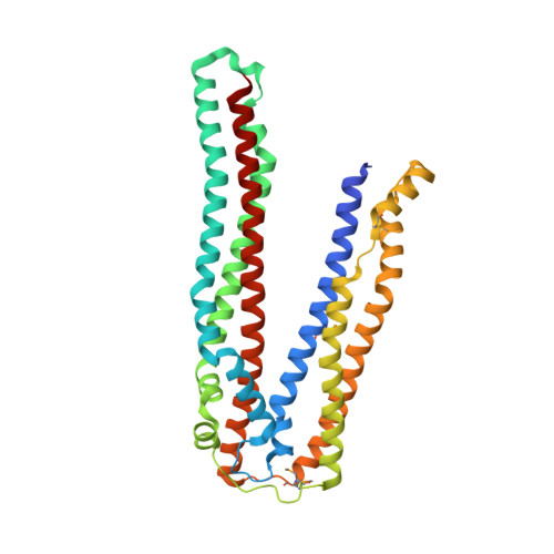
    ALIX/AIP1 functions in enveloped virus budding, endosomal protein sorting, and many other cellular processes. Retroviruses, including HIV-1, SIV, and EIAV, bind and recruit ALIX through YPX(n)L late-domain motifs (X = any residue; n = 1-3). Crystal structures reveal that human ALIX is composed of an N-terminal Bro1 domain and a central domain that is composed of two extended three-helix bundles that form elongated arms that fold back into a "V." The structures also reveal conformational flexibility in the arms that suggests that the V domain may act as a flexible hinge in response to ligand binding. YPX(n)L late domains bind in a conserved hydrophobic pocket on the second arm near the apex of the V, whereas CHMP4/ESCRT-III proteins bind a conserved hydrophobic patch on the Bro1 domain, and both interactions are required for virus budding. ALIX therefore serves as a flexible, extended scaffold that connects retroviral Gag proteins to ESCRT-III and other cellular-budding machinery.


  • Organizational Affiliation

    Department of Biochemistry, University of Utah School of Medicine, Salt Lake City, UT 84112, USA.


Macromolecules
Find similar proteins by:  (by identity cutoff)  |  3D Structure
Entity ID: 1
MoleculeChains Sequence LengthOrganismDetailsImage
Programmed cell death 6-interacting protein
A, B
351Homo sapiensMutation(s): 4 
Gene Names: PDCD6IPAIP1ALIXKIAA1375
UniProt & NIH Common Fund Data Resources
Find proteins for Q8WUM4 (Homo sapiens)
Explore Q8WUM4 
Go to UniProtKB:  Q8WUM4
PHAROS:  Q8WUM4
GTEx:  ENSG00000170248 
Entity Groups  
Sequence Clusters30% Identity50% Identity70% Identity90% Identity95% Identity100% Identity
UniProt GroupQ8WUM4
Sequence Annotations
Expand
  • Reference Sequence
Small Molecules
Modified Residues  1 Unique
IDChains TypeFormula2D DiagramParent
MSE
Query on MSE
A, B
L-PEPTIDE LINKINGC5 H11 N O2 SeMET
Experimental Data & Validation

Experimental Data

  • Method: X-RAY DIFFRACTION
  • Resolution: 2.58 Å
  • R-Value Free: 0.302 
  • R-Value Work: 0.228 
  • R-Value Observed: 0.231 
  • Space Group: P 1 21 1
Unit Cell:
Length ( Å )Angle ( ˚ )
a = 63.861α = 90
b = 50.049β = 101.13
c = 130.171γ = 90
Software Package:
Software NamePurpose
DENZOdata reduction
SCALEPACKdata scaling
SOLVEphasing
RESOLVEphasing
REFMACrefinement
PDB_EXTRACTdata extraction
ADSCdata collection
HKL-2000data reduction

Structure Validation

View Full Validation Report


Entry History 

Deposition Data

Revision History  (Full details and data files)

  • Version 1.0: 2007-03-27
    Type: Initial release
  • Version 1.1: 2008-05-01
    Changes: Version format compliance
  • Version 1.2: 2011-07-13
    Changes: Version format compliance
  • Version 1.3: 2017-10-18
    Changes: Refinement description
  • Version 1.4: 2023-12-27
    Changes: Data collection, Database references, Derived calculations