2O3O | pdb_00002o3o

Crystal Structure of the sensor histidine kinase regulator YycI from Bacillus subtitlis


Experimental Data Snapshot

  • Method: X-RAY DIFFRACTION
  • Resolution: 2.89 Å
  • R-Value Free: 
    0.262 (Depositor), 0.294 (DCC) 
  • R-Value Work: 
    0.212 (Depositor), 0.254 (DCC) 
  • R-Value Observed: 
    0.214 (Depositor) 

wwPDB Validation 3D Report Full Report

Validation slider image for 2O3O

This is version 1.4 of the entry. See complete history

Literature

The Crystal Structure of Bacillus subtilis YycI Reveals a Common Fold for Two Members of an Unusual Class of Sensor Histidine Kinase Regulatory Proteins.

Santelli, E.Liddington, R.C.Mohan, M.A.Hoch, J.A.Szurmant, H.

(2007) J Bacteriol 189: 3290-3295

  • DOI: https://doi.org/10.1128/JB.01937-06
  • Primary Citation Related Structures: 
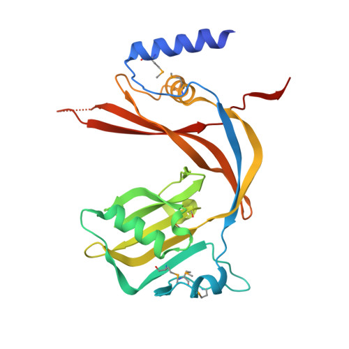
    2O3O

  • PubMed Abstract: 

    YycI and YycH are two membrane-anchored periplasmic proteins that regulate the essential Bacillus subtilis YycG histidine kinase through direct interaction. Here we present the crystal structure of YycI at a 2.9-A resolution. YycI forms a dimer, and remarkably the structure resembles that of the two C-terminal domains of YycH despite nearly undetectable sequence homology (10%) between the two proteins.


  • Organizational Affiliation
    • Division of Cellular Biology, Mail code MEM-116, Department of Molecular and Experimental Medicine, The Scripps Research Institute, 10550 North Torrey Pines Road, La Jolla, CA 92037, USA.

Macromolecule Content 

  • Total Structure Weight: 355.85 kDa 
  • Atom Count: 23,401 
  • Modeled Residue Count: 2,871 
  • Deposited Residue Count: 3,048 
  • Unique protein chains: 1

Macromolecules

Find similar proteins by:|  3D Structure
Entity ID: 1
MoleculeChains  Sequence LengthOrganismDetailsImage
YycI protein
A, B, C, D, E
A, B, C, D, E, F, G, H, I, J, K, L
254Bacillus subtilisMutation(s): 6 
Gene Names: yycI
UniProt
Find proteins for Q45612 (Bacillus subtilis (strain 168))
Explore Q45612 
Go to UniProtKB:  Q45612
Entity Groups
Sequence Clusters30% Identity50% Identity70% Identity90% Identity95% Identity100% Identity
UniProt GroupQ45612
Sequence Annotations
Expand
Reference Sequence

Experimental Data & Validation

Experimental Data

  • Method: X-RAY DIFFRACTION
  • Resolution: 2.89 Å
  • R-Value Free:  0.262 (Depositor), 0.294 (DCC) 
  • R-Value Work:  0.212 (Depositor), 0.254 (DCC) 
  • R-Value Observed: 0.214 (Depositor) 
Space Group: P 1 21 1
Unit Cell:
Length ( Å )Angle ( ˚ )
a = 60.792α = 90
b = 161.922β = 90.9
c = 180.156γ = 90
Software Package:
Software NamePurpose
REFMACrefinement
HKL-2000data reduction
HKL-2000data scaling
SHELXDphasing
SHARPphasing

Structure Validation

View Full Validation Report


Entry History 

Deposition Data

Revision History  (Full details and data files)

  • Version 1.0: 2007-04-10
    Type: Initial release
  • Version 1.1: 2008-05-01
    Changes: Version format compliance
  • Version 1.2: 2011-07-13
    Changes: Advisory, Refinement description, Version format compliance
  • Version 1.3: 2023-12-27
    Changes: Data collection, Database references, Derived calculations, Refinement description
  • Version 1.4: 2024-10-16
    Changes: Structure summary