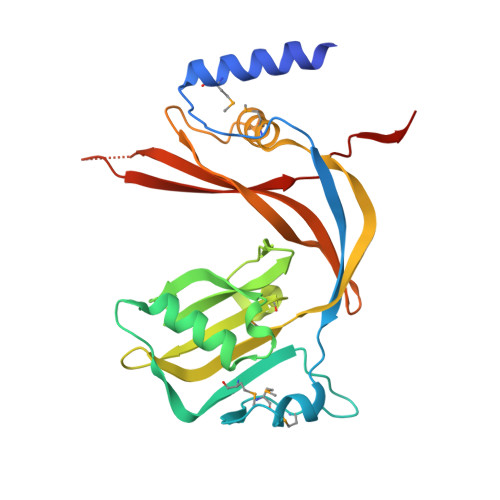
The Crystal Structure of Bacillus subtilis YycI Reveals a Common Fold for Two Members of an Unusual Class of Sensor Histidine Kinase Regulatory Proteins.
Santelli, E., Liddington, R.C., Mohan, M.A., Hoch, J.A., Szurmant, H.(2007) J Bacteriol 189: 3290-3295
- PubMed: 17307848
- DOI: https://doi.org/10.1128/JB.01937-06
- Primary Citation of Related Structures:
2O3O - PubMed Abstract:
YycI and YycH are two membrane-anchored periplasmic proteins that regulate the essential Bacillus subtilis YycG histidine kinase through direct interaction. Here we present the crystal structure of YycI at a 2.9-A resolution. YycI forms a dimer, and remarkably the structure resembles that of the two C-terminal domains of YycH despite nearly undetectable sequence homology (10%) between the two proteins.
- Division of Cellular Biology, Mail code MEM-116, Department of Molecular and Experimental Medicine, The Scripps Research Institute, 10550 North Torrey Pines Road, La Jolla, CA 92037, USA.
Organizational Affiliation:


