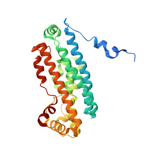
2NOX

Crystal structure of tryptophan 2,3-dioxygenase from Ralstonia metallidurans


Experimental Data Snapshot

  • Method: X-RAY DIFFRACTION
  • Resolution: 2.40 Å
  • R-Value Free: 0.270 
  • R-Value Work: 0.210 
  • R-Value Observed: 0.210 

Starting Model: experimental
View more details

wwPDB Validation   3D Report Full Report


Ligand Structure Quality Assessment 


This is version 1.4 of the entry. See complete history


Literature

Crystal structure and mechanism of tryptophan 2,3-dioxygenase, a heme enzyme involved in tryptophan catabolism and in quinolinate biosynthesis.

Zhang, Y.Kang, S.A.Mukherjee, T.Bale, S.Crane, B.R.Begley, T.P.Ealick, S.E.

(2007) Biochemistry 46: 145-155

  • DOI: https://doi.org/10.1021/bi0620095
  • Primary Citation of Related Structures:  
    2NOX

  • PubMed Abstract: 

    The structure of tryptophan 2,3-dioxygenase (TDO) from Ralstonia metallidurans was determined at 2.4 A. TDO catalyzes the irreversible oxidation of l-tryptophan to N-formyl kynurenine, which is the initial step in tryptophan catabolism. TDO is a heme-containing enzyme and is highly specific for its substrate l-tryptophan. The structure is a tetramer with a heme cofactor bound at each active site. The monomeric fold, as well as the heme binding site, is similar to that of the large domain of indoleamine 2,3-dioxygenase, an enzyme that catalyzes the same reaction except with a broader substrate tolerance. Modeling of the putative (S)-tryptophan hydroperoxide intermediate into the active site, as well as substrate analogue and mutagenesis studies, are consistent with a Criegee mechanism for the reaction.


  • Organizational Affiliation

    Department of Chemistry and Chemical Biology, Cornell University, Ithaca, New York 14853, USA.


Macromolecules
Find similar proteins by:  (by identity cutoff)  |  3D Structure
Entity ID: 1
MoleculeChains Sequence LengthOrganismDetailsImage
Tryptophan 2,3-dioxygenase
A, B, C, D, E
A, B, C, D, E, F, G, H, I, J, K, L, M, N, O, P
281Cupriavidus metalliduransMutation(s): 0 
EC: 1.13.11.11
UniProt
Find proteins for Q1LK00 (Cupriavidus metallidurans (strain ATCC 43123 / DSM 2839 / NBRC 102507 / CH34))
Explore Q1LK00 
Go to UniProtKB:  Q1LK00
Entity Groups  
Sequence Clusters30% Identity50% Identity70% Identity90% Identity95% Identity100% Identity
UniProt GroupQ1LK00
Sequence Annotations
Expand
  • Reference Sequence
Small Molecules
Ligands 1 Unique
IDChains Name / Formula / InChI Key2D Diagram3D Interactions
HEM
Query on HEM

Download Ideal Coordinates CCD File 
AA [auth K]
BA [auth L]
CA [auth M]
DA [auth N]
EA [auth O]
AA [auth K],
BA [auth L],
CA [auth M],
DA [auth N],
EA [auth O],
FA [auth P],
Q [auth A],
R [auth B],
S [auth C],
T [auth D],
U [auth E],
V [auth F],
W [auth G],
X [auth H],
Y [auth I],
Z [auth J]
PROTOPORPHYRIN IX CONTAINING FE
C34 H32 Fe N4 O4
KABFMIBPWCXCRK-RGGAHWMASA-L
Experimental Data & Validation

Experimental Data

  • Method: X-RAY DIFFRACTION
  • Resolution: 2.40 Å
  • R-Value Free: 0.270 
  • R-Value Work: 0.210 
  • R-Value Observed: 0.210 
  • Space Group: P 1
Unit Cell:
Length ( Å )Angle ( ˚ )
a = 72.544α = 66.97
b = 132.121β = 85.06
c = 139.95γ = 89.89
Software Package:
Software NamePurpose
DENZOdata reduction
SCALEPACKdata scaling
REFMACrefinement
PDB_EXTRACTdata extraction
ADSCdata collection
HKL-2000data reduction
CNSphasing

Structure Validation

View Full Validation Report


Ligand Structure Quality Assessment 


Entry History 

Deposition Data

Revision History  (Full details and data files)

  • Version 1.0: 2006-12-19
    Type: Initial release
  • Version 1.1: 2008-05-01
    Changes: Version format compliance
  • Version 1.2: 2011-07-13
    Changes: Advisory, Version format compliance
  • Version 1.3: 2017-10-18
    Changes: Refinement description
  • Version 1.4: 2023-08-30
    Changes: Data collection, Database references, Derived calculations, Refinement description