Crystal structure and mechanism of tryptophan 2,3-dioxygenase, a heme enzyme involved in tryptophan catabolism and in quinolinate biosynthesis.
Zhang, Y., Kang, S.A., Mukherjee, T., Bale, S., Crane, B.R., Begley, T.P., Ealick, S.E.(2007) Biochemistry 46: 145-155
- PubMed: 17198384 Search on PubMed
- DOI: https://doi.org/10.1021/bi0620095
- Primary Citation Related Structures:
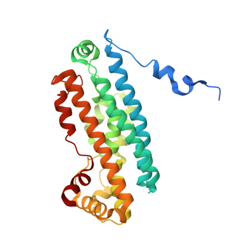
2NOX - PubMed Abstract:
The structure of tryptophan 2,3-dioxygenase (TDO) from Ralstonia metallidurans was determined at 2.4 A. TDO catalyzes the irreversible oxidation of l-tryptophan to N-formyl kynurenine, which is the initial step in tryptophan catabolism. TDO is a heme-containing enzyme and is highly specific for its substrate l-tryptophan. The structure is a tetramer with a heme cofactor bound at each active site. The monomeric fold, as well as the heme binding site, is similar to that of the large domain of indoleamine 2,3-dioxygenase, an enzyme that catalyzes the same reaction except with a broader substrate tolerance. Modeling of the putative (S)-tryptophan hydroperoxide intermediate into the active site, as well as substrate analogue and mutagenesis studies, are consistent with a Criegee mechanism for the reaction.
- Department of Chemistry and Chemical Biology, Cornell University, Ithaca, New York 14853, USA.
Organizational Affiliation: