2MRU | pdb_00002mru

Structure of truncated EcMazE-DNA complex


Experimental Data Snapshot

  • Method: SOLUTION NMR
  • Conformers Calculated: 100 
  • Conformers Submitted: 
  • Selection Criteria: structures with the least restraint violations 

wwPDB Validation 3D Report Full Report

Validation slider image for 2MRU

This is version 1.2 of the entry. See complete history

Literature

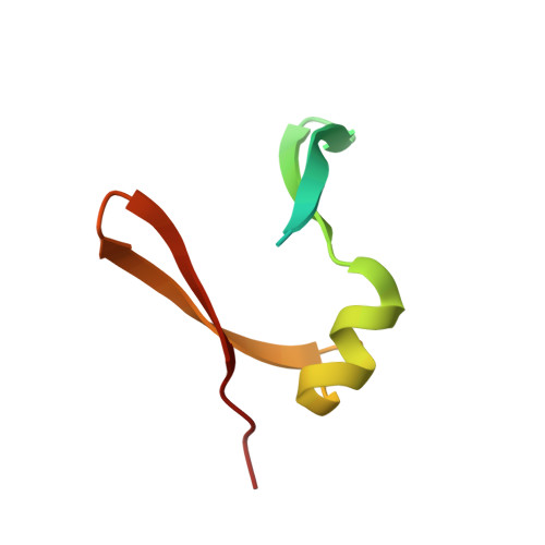
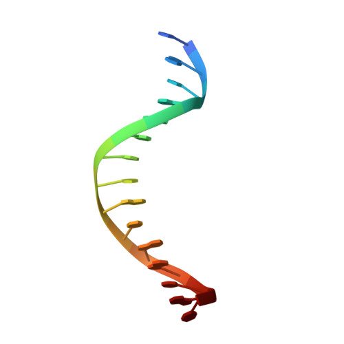
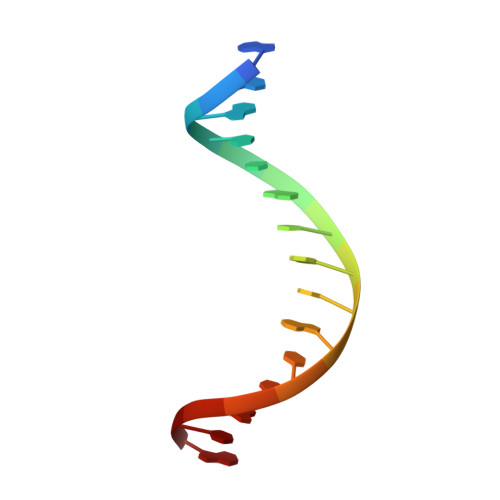
Escherichia coli antitoxin MazE as transcription factor: insights into MazE-DNA binding.

Zorzini, V.Buts, L.Schrank, E.Sterckx, Y.G.Respondek, M.Engelberg-Kulka, H.Loris, R.Zangger, K.van Nuland, N.A.

(2015) Nucleic Acids Res 43: 1241-1256

  • DOI: https://doi.org/10.1093/nar/gku1352
  • Primary Citation Related Structures: 
    2MRN, 2MRU

  • PubMed Abstract: 

    Toxin-antitoxin (TA) modules are pairs of genes essential for bacterial regulation upon environmental stresses. The mazEF module encodes the MazF toxin and its cognate MazE antitoxin. The highly dynamic MazE possesses an N-terminal DNA binding domain through which it can negatively regulate its own promoter. Despite being one of the first TA systems studied, transcriptional regulation of Escherichia coli mazEF remains poorly understood. This paper presents the solution structure of C-terminal truncated E. coli MazE and a MazE-DNA model with a DNA palindrome sequence ∼ 10 bp upstream of the mazEF promoter. The work has led to a transcription regulator-DNA model, which has remained elusive thus far in the E. coli toxin-antitoxin family. Multiple complementary techniques including NMR, SAXS and ITC show that the long intrinsically disordered C-termini in MazE, required for MazF neutralization, does not affect the interactions between the antitoxin and its operator. Rather, the MazE C-terminus plays an important role in the MazF binding, which was found to increase the MazE affinity for the palindromic single site operator.


  • Organizational Affiliation
    • Molecular Recognition Unit, Structural Biology Research Center, VIB, Pleinlaan 2, 1050 Brussels, Belgium Structural Biology Brussels, Vrije Universiteit Brussel, Pleinlaan 2, 1050 Brussels, Belgium.

Macromolecule Content 

  • Total Structure Weight: 24.53 kDa 
  • Atom Count: 1,392 
  • Modeled Residue Count: 130 
  • Deposited Residue Count: 164 
  • Unique protein chains: 1
  • Unique nucleic acid chains: 2

Macromolecules


Find similar proteins by:|  3D Structure
Entity ID: 1
MoleculeChains  Sequence LengthOrganismDetailsImage
Antitoxin MazE
A, B
67Escherichia coli K-12Mutation(s): 0 
Gene Names: b2783BN896_2518chpAIchpRJW2754mazE
UniProt
Find proteins for P0AE72 (Escherichia coli (strain K12))
Explore P0AE72 
Go to UniProtKB:  P0AE72
Entity Groups
Sequence Clusters30% Identity50% Identity70% Identity90% Identity95% Identity100% Identity
UniProt GroupP0AE72
Sequence Annotations
Expand
Reference Sequence
Find similar nucleic acids by:  Sequence
Entity ID: 2
MoleculeChains LengthOrganismImage
DNA (5'-D(*CP*GP*TP*GP*AP*TP*AP*TP*AP*TP*AP*GP*TP*GP*C)-3')C [auth X]15N/A
Sequence Annotations
Expand
Reference Sequence
Find similar nucleic acids by:  Sequence
Entity ID: 3
MoleculeChains LengthOrganismImage
DNA (5'-D(P*GP*CP*AP*CP*TP*AP*TP*AP*TP*AP*TP*CP*AP*CP*G)-3')D [auth Y]15N/A
Sequence Annotations
Expand
Reference Sequence

Experimental Data & Validation

Experimental Data

  • Method: SOLUTION NMR
  • Conformers Calculated: 100 
  • Conformers Submitted: 
  • Selection Criteria: structures with the least restraint violations 

Structure Validation

View Full Validation Report


Entry History 

Deposition Data

Revision History  (Full details and data files)

  • Version 1.0: 2015-02-04
    Type: Initial release
  • Version 1.1: 2023-06-14
    Changes: Data collection, Database references, Other
  • Version 1.2: 2024-05-15
    Changes: Data collection, Database references