2MAD

THE ACTIVE SITE STRUCTURE OF METHYLAMINE DEHYDROGENASE: HYDRAZINES IDENTIFY C6 AS THE REACTIVE SITE OF THE TRYPTOPHAN DERIVED QUINONE COFACTOR


Experimental Data Snapshot

  • Method: X-RAY DIFFRACTION
  • Resolution: 2.25 Å
  • R-Value Observed: 0.209 

wwPDB Validation   3D Report Full Report


This is version 1.2 of the entry. See complete history


Literature

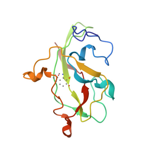
Active site structure of methylamine dehydrogenase: hydrazines identify C6 as the reactive site of the tryptophan-derived quinone cofactor.

Huizinga, E.G.van Zanten, B.A.Duine, J.A.Jongejan, J.A.Huitema, F.Wilson, K.S.Hol, W.G.

(1992) Biochemistry 31: 9789-9795

  • DOI: 10.1021/bi00155a036
  • Primary Citation of Related Structures:  
    1MAE, 1MAF, 2MAD

  • PubMed Abstract: 
  • To identify the reactive part of the orthoquinone function of the tryptophan-derived cofactor found in methylamine dehydrogenase (MADH), we have determined the crystal structures of MADH from Thiobacillus versutus inhibited by methylhydrazine and (2,2,2-trifluoroethyl)hydrazine ...

    To identify the reactive part of the orthoquinone function of the tryptophan-derived cofactor found in methylamine dehydrogenase (MADH), we have determined the crystal structures of MADH from Thiobacillus versutus inhibited by methylhydrazine and (2,2,2-trifluoroethyl)hydrazine. Extra electron density attached to C6 of the tryptophyl tryptophanquinone cofactor shows that this atom and not C7 is the reactive part of the ortho-quinone moiety. The density retained after hydrazine inhibition is much less extensive than expected, however, suggesting that partial breakdown of the inhibitors after reaction with the cofactor may take place. A detailed description is presented of the cofactor environment in an improved model of MADH which now includes information from the recently determined gene sequence of the cofactor-containing subunit [Ubbink, M., van Kleef, M.A.G., Kleinjan, D., Hoitink, C.W.G., Huitema, F., Beintema, J.J., Duine, J.A., & Canters, G.W. (1991) Eur. J. Biochem. 202, 1003-1012]. We hypothesize that Asp76 is responsible for proton abstraction from the alpha-carbon of the substrate during catalysis.


    Related Citations: 
    • Crystallographic Investigations of the Tryptophan-Derived Cofactor in the Quinoprotein Methylamine Dehydrogenase
      Chen, L., Mathews, F.S., Davidson, V.L., Huizinga, E.G., Vellieux, F.M.D., Duine, J.A., Hol, W.G.J.
      (1991) FEBS Lett 287: 163
    • Structure of Quinoprotein Methylamine Dehydrogenase at 2.25 Angstroms Resolution
      Vellieux, F.M.D., Huitema, F., Groendijk, H., Kalk, K.H., Frank, J., Jongejan, J.A., Duine, J.A., Petratos, K., Drenth, J., Hol, W.G.J.
      (1989) EMBO J 8: 2171
    • Purification, Crystallization and Preliminary X-Ray Investigation of Quinoprotein Methylamine Dehydrogenase from Thiobacillius Versutus
      Vellieux, F.M.D., Frank, J., Swarte, M.B.A., Groendijk, H., Duine, J.A., Drenth, J., Hol, W.G.J.
      (1986) Eur J Biochem 154: 383

    Organizational Affiliation

    Department of Chemistry, Groningen University, The Netherlands.


Macromolecules
Find similar proteins by:  (by identity cutoff)  |  3D Structure
Entity ID: 1
MoleculeChainsSequence LengthOrganismDetailsImage
METHYLAMINE DEHYDROGENASE (LIGHT SUBUNIT)A [auth L]124Paracoccus versutusMutation(s): 0 
Gene Names: mauAmadB
EC: 1.4.99.3 (PDB Primary Data), 1.4.9.1 (UniProt)
UniProt
Find proteins for P22641 (Paracoccus versutus)
Explore P22641 
Go to UniProtKB:  P22641
Entity Groups  
Sequence Clusters30% Identity50% Identity70% Identity90% Identity95% Identity100% Identity
UniProt GroupP22641
Protein Feature View
Expand
  • Reference Sequence
Find similar proteins by:  (by identity cutoff)  |  3D Structure
Entity ID: 2
MoleculeChainsSequence LengthOrganismDetailsImage
METHYLAMINE DEHYDROGENASE (HEAVY SUBUNIT)B [auth H]373Paracoccus versutusMutation(s): 0 
Gene Names: mauBmadA
EC: 1.4.99.3 (PDB Primary Data), 1.4.9.1 (UniProt)
UniProt
Find proteins for P23006 (Paracoccus versutus)
Explore P23006 
Go to UniProtKB:  P23006
Entity Groups  
Sequence Clusters30% Identity50% Identity70% Identity90% Identity95% Identity100% Identity
UniProt GroupP23006
Protein Feature View
Expand
  • Reference Sequence
Small Molecules
Modified Residues  1 Unique
IDChainsTypeFormula2D DiagramParent
TRQ
Query on TRQ
A [auth L]L-PEPTIDE LINKINGC11 H10 N2 O4TRP
Experimental Data & Validation

Experimental Data

  • Method: X-RAY DIFFRACTION
  • Resolution: 2.25 Å
  • R-Value Observed: 0.209 
  • Space Group: P 31 2 1
Unit Cell:
Length ( Å )Angle ( ˚ )
a = 129.784α = 90
b = 129.784β = 90
c = 104.334γ = 120
Software Package:
Software NamePurpose
TNTrefinement

Structure Validation

View Full Validation Report


Entry History 

Deposition Data

Revision History  (Full details and data files)

  • Version 1.0: 1994-01-31
    Type: Initial release
  • Version 1.1: 2008-03-24
    Changes: Version format compliance
  • Version 1.2: 2011-07-13
    Changes: Version format compliance