Structural characterization of AS1-membrane interactions from a subset of HAMP domains
Unnerstale, S., Maler, L., Draheim, R.R.(2011) Biochim Biophys Acta 1808: 2403-2412
- PubMed: 21763270 Search on PubMedSearch on PubMed Central
- DOI: https://doi.org/10.1016/j.bbamem.2011.06.018
- Primary Citation Related Structures:
2L9G - PubMed Abstract:
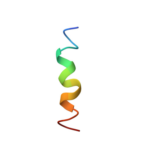
HAMP domains convert an extracellular sensory input into an intracellular signaling response in a wide variety of membrane-embedded bacterial proteins. These domains are almost invariably found adjacent to the inner leaflet of the cell membrane. We therefore examined the interaction of peptides corresponding to either AS1 or AS2 of four different, well-characterized HAMP domains with several membrane model systems. The proteins included an Archaeoglobus fulgidus protein (Af1503), the Escherichia coli osmosensor EnvZ(Ec), the E. coli nitrate/nitrite sensor NarX(Ec), and the aspartate chemoreceptor of E. coli (Tar(Ec)). Far-UV CD and NMR spectroscopy were used to monitor the induction of secondary structure upon association with neutral or acidic large unilamellar vesicles (LUVs) and bicelles. We observed significant increases in α-helicity within AS1 from NarX(Ec) and Tar(Ec) but not in AS1 from the other proteins. To characterize these interactions further, we determined the solution structure of AS1 from Tar(Ec) associated with acidic bicelles. The bulk of AS1 formed an amphipathic α-helix, whereas the N-terminal control cable, the region between TM2 and AS1, remained unstructured. We observed that the conserved prolyl residue found in AS1 of many membrane-adjacent HAMP domains defined the boundary between the unstructured and helical regions. In addition, two positively charged residues that flank the hydrophobic surface of AS1 are thought to facilitate electrostatic interactions with the membrane. We interpret these results within the context of the helix-interaction model for HAMP signaling and propose roles for AS1-membrane interactions during the membrane assembly and transmembrane communication of HAMP-containing receptors.
- Department of Biochemistry and Biophysics, The Arrhenius Laboratories for Natural Sciences, Stockholm University, Stockholm, Sweden.
Organizational Affiliation: