NMR structural characterization of HIV-1 virus protein U cytoplasmic domain in the presence of dodecylphosphatidylcholine micelles
Wittlich, M., Koenig, B.W., Stoldt, M., Schmidt, H., Willbold, D.(2009) FEBS J 276: 6560-6575
- PubMed: 19804408 Search on PubMed
- DOI: https://doi.org/10.1111/j.1742-4658.2009.07363.x
- Primary Citation Related Structures:
2K7Y - PubMed Abstract:
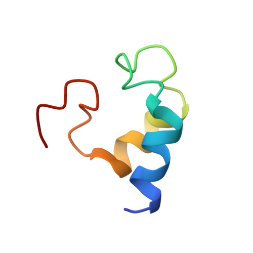
The HIV-1 encoded virus protein U (VpU) is required for efficient viral release from human host cells and for induction of CD4 degradation in the endoplasmic reticulum. The cytoplasmic domain of the membrane protein VpU (VpUcyt) is essential for the latter activity. The structure and dynamics of VpUcyt were characterized in the presence of membrane simulating dodecylphosphatidylcholine (DPC) micelles by high-resolution liquid state NMR. VpUcyt is unstructured in aqueous buffer. The addition of DPC micelles induces a well-defined membrane proximal alpha-helix (residues I39-E48) and an additional helical segment (residues L64-R70). A tight loop (L73-V78) is observed close to the C-terminus, whereas the interhelical linker (R49-E63) remains highly flexible. A 3D structure of VpUcyt in the presence of DPC micelles was calculated from a large set of proton-proton distance constraints. The topology of micelle-associated VpUcyt was derived from paramagnetic relaxation enhancement of protein nuclear spins after the introduction of paramagnetic probes into the interior of the micelle or the aqueous buffer. Qualitative analysis of secondary chemical shift and paramagnetic relaxation enhancement data in conjunction with dynamic information from heteronuclear NOEs and structural insight from homonuclear NOE-based distance constraints indicated that micelle-associated VpUcyt retains a substantial degree of structural flexibility.
- Institut für Strukturbiologie und Biophysik (ISB-3), Forschungszentrum Jülich, Germany.
Organizational Affiliation: