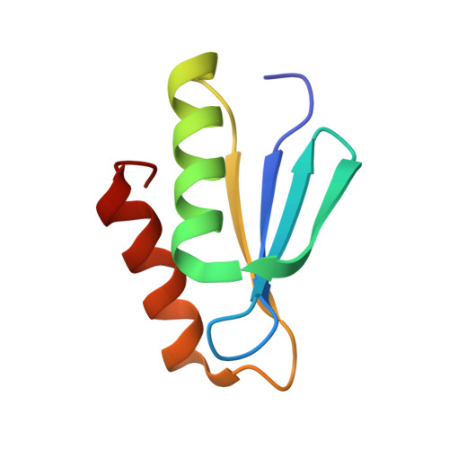
The Periplasmic Domain of TolR from Haemophilus influenzae Forms a Dimer with a Large Hydrophobic Groove: NMR Solution Structure and Comparison to SAXS Data.
Parsons, L.M., Grishaev, A., Bax, A.(2008) Biochemistry 47: 3131-3142
- PubMed: 18269247 Search on PubMed
- DOI: https://doi.org/10.1021/bi702283x
- Primary Citation Related Structures:
2JWK, 2JWL - PubMed Abstract:
TolR is a part of the Pal/Tol system which forms a five-member, membrane-spanning, multiprotein complex that is conserved in Gram-negative bacteria. The Pal/Tol system helps to maintain the integrity of the outer membrane and has been proposed to be involved in several other cellular processes including cell division. Obtaining the structure of TolR is of interest not only to help explain the many proposed functions of the Pal/Tol system but also to gain an understanding of the TolR homologues ExbD and MotB and to provide more targets for antibacterial treatments. In addition, the structure may provide insights into how colicins and bacteriophages are able to enter the cell. Here we report the solution structure of the homodimeric periplasmic domain of TolR from Haemophilus influenzae, determined with conventional, NOE-based NMR spectroscopy, supplemented by extensive residual dipolar coupling measurements. A novel method for assembling the dimer from small-angle X-ray scattering data confirms the NMR-derived structure. To facilitate NMR spectral analysis, a TolR construct containing residues 59-130 of the 139-residue protein was created. The periplasmic domain of TolR forms a C 2-symmetric dimer consisting of a strongly curved eight-stranded beta-sheet, generating a large deep groove on one side, while four helices cover the other face of the sheet. The structure of the TolR dimer together with data from the literature suggests how the periplasmic domain of TolR is most likely oriented relative to the cytoplasmic membrane and how it may interact with other components of the Pal/Tol system, particularly TolQ.
- Laboratory of Chemical Physics, National Institute of Diabetes and Digestive and Kidney Diseases, National Institutes of Health, 5 Memorial Drive, Bethesda, Maryland 20892, USA.
Organizational Affiliation: