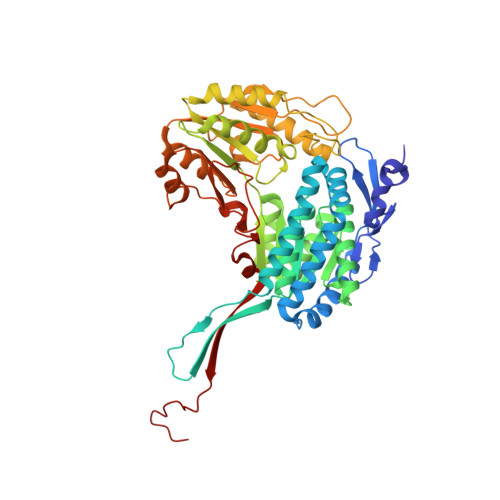
2JG7

Crystal structure of Seabream Antiquitin and Elucidation of its substrate specificity


Experimental Data Snapshot

  • Method: X-RAY DIFFRACTION
  • Resolution: 2.83 Å
  • R-Value Free: 0.228 
  • R-Value Work: 0.210 
  • R-Value Observed: 0.211 

Starting Model: experimental
View more details

wwPDB Validation   3D Report Full Report


Ligand Structure Quality Assessment 


This is version 1.3 of the entry. See complete history


Literature

The Crystal Structure of Seabream Antiquitin Reveals the Structural Basis of its Substrate Specificity.

Tang, W.K.Wong, K.B.Lam, Y.M.Cha, S.S.Cheng, C.H.K.Fong, W.P.

(2008) FEBS Lett 582: 3090

  • DOI: https://doi.org/10.1016/j.febslet.2008.07.059
  • Primary Citation of Related Structures:  
    2JG7

  • PubMed Abstract: 

    The crystal structure of seabream antiquitin in complex with the cofactor NAD(+) was solved at 2.8A resolution. The mouth of the substrate-binding pocket is guarded by two conserved residues, Glu120 and Arg300. To test the role of these two residues, we have prepared the two mutants E120A and R300A. Our model and kinetics data suggest that antiquitin's specificity towards the substrate alpha-aminoadipic semialdehyde is contributed mainly by Glu120 which interacts with the alpha-amino group of the substrate. On the other hand, Arg300 does not have any specific interaction with the alpha-carboxylate group of the substrate, but is important in maintaining the active site conformation.


  • Organizational Affiliation

    Department of Biochemistry, The Chinese University of Hong Kong, Shatin, N.T., Hong Kong, China.


Macromolecules
Find similar proteins by:  (by identity cutoff)  |  3D Structure
Entity ID: 1
MoleculeChains Sequence LengthOrganismDetailsImage
ANTIQUITIN
A, B, C, D, E
A, B, C, D, E, F, G, H
510Acanthopagrus schlegeliiMutation(s): 0 
UniProt
Find proteins for Q4KTQ7 (Acanthopagrus schlegelii)
Explore Q4KTQ7 
Go to UniProtKB:  Q4KTQ7
Entity Groups  
Sequence Clusters30% Identity50% Identity70% Identity90% Identity95% Identity100% Identity
UniProt GroupQ4KTQ7
Sequence Annotations
Expand
  • Reference Sequence
Experimental Data & Validation

Experimental Data

  • Method: X-RAY DIFFRACTION
  • Resolution: 2.83 Å
  • R-Value Free: 0.228 
  • R-Value Work: 0.210 
  • R-Value Observed: 0.211 
  • Space Group: P 1
Unit Cell:
Length ( Å )Angle ( ˚ )
a = 88.16α = 90.5
b = 111.02β = 98.1
c = 113.64γ = 111.59
Software Package:
Software NamePurpose
REFMACrefinement

Structure Validation

View Full Validation Report


Ligand Structure Quality Assessment 


Entry History 

Deposition Data

Revision History  (Full details and data files)

  • Version 1.0: 2008-05-13
    Type: Initial release
  • Version 1.1: 2011-05-08
    Changes: Version format compliance
  • Version 1.2: 2011-07-13
    Changes: Version format compliance
  • Version 1.3: 2023-12-13
    Changes: Data collection, Database references, Other, Refinement description