2J12

Ad37 fibre head in complex with CAR D1


Experimental Data Snapshot

  • Method: X-RAY DIFFRACTION
  • Resolution: 1.50 Å
  • R-Value Free: 0.169 
  • R-Value Work: 0.148 
  • R-Value Observed: 0.149 

wwPDB Validation   3D Report Full Report


This is version 2.0 of the entry. See complete history


Literature

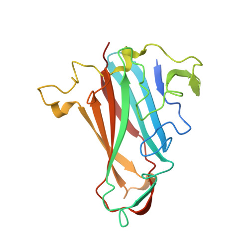
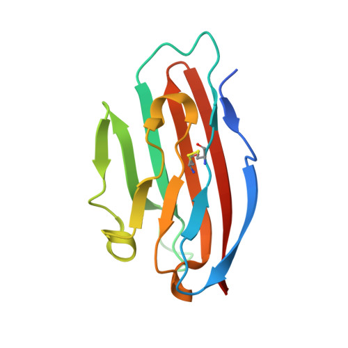
Structural and Mutational Analysis of Human Ad37 and Canine Adenovirus 2 Fiber Heads in Complex with the D1 Domain of Coxsackie and Adenovirus Receptor.

Seiradake, E.Lortat-Jacob, H.Billet, O.Kremer, E.J.Cusack, S.

(2006) J Biol Chem 281: 33704

  • DOI: https://doi.org/10.1074/jbc.M605316200
  • Primary Citation of Related Structures:  
    2J12, 2J1K, 2J2J

  • PubMed Abstract: 
  • Adenovirus fibers from most serotypes bind the D1 domain of coxsackie and adenovirus receptor (CAR), although the binding residues are not strictly conserved. To understand this further, we determined the crystal structures of canine adenovirus serotype 2 (CAV-2) and the human adenovirus serotype 37 (HAd37) in complex with human CAR D1 at 2 ...

    Adenovirus fibers from most serotypes bind the D1 domain of coxsackie and adenovirus receptor (CAR), although the binding residues are not strictly conserved. To understand this further, we determined the crystal structures of canine adenovirus serotype 2 (CAV-2) and the human adenovirus serotype 37 (HAd37) in complex with human CAR D1 at 2.3 and 1.5A resolution, respectively. Structure comparison with the HAd12 fiber head-CAR D1 complex showed that the overall topology of the interaction is conserved but that the interfaces differ in number and identity of interacting residues, shape complementarity, and degree of conformational adaptation. Using surface plasmon resonance, we characterized the binding affinity to CAR D1 of wild type and mutant CAV-2 and HAd37 fiber heads. We found that CAV-2 has the highest affinity but fewest direct interactions, with the reverse being true for HAd37. Moreover, we found that conserved interactions can have a minor contribution, whereas serotype-specific interactions can be essential. These results are discussed in the light of virus evolution and design of adenovirus vectors for gene transfer.


    Organizational Affiliation

    Grenoble Outstation, European Molecular Biology Laboratory, Grenoble, France.


Macromolecules
Find similar proteins by:  (by identity cutoff)  |  3D Structure
Entity ID: 1
MoleculeChainsSequence LengthOrganismDetailsImage
FIBER PROTEINA194Human adenovirus D37Mutation(s): 0 
UniProt
Find proteins for Q80S15 (Human adenovirus D37)
Explore Q80S15 
Go to UniProtKB:  Q80S15
Entity Groups  
Sequence Clusters30% Identity50% Identity70% Identity90% Identity95% Identity100% Identity
UniProt GroupQ80S15
Protein Feature View
Expand
  • Reference Sequence
Find similar proteins by:  (by identity cutoff)  |  3D Structure
Entity ID: 2
MoleculeChainsSequence LengthOrganismDetailsImage
COXSACKIEVIRUS AND ADENOVIRUS RECEPTORB128Homo sapiensMutation(s): 0 
Gene Names: CXADRCAR
UniProt & NIH Common Fund Data Resources
Find proteins for P78310 (Homo sapiens)
Explore P78310 
Go to UniProtKB:  P78310
PHAROS:  P78310
Entity Groups  
Sequence Clusters30% Identity50% Identity70% Identity90% Identity95% Identity100% Identity
UniProt GroupP78310
Protein Feature View
Expand
  • Reference Sequence
Small Molecules
Ligands 1 Unique
IDChainsName / Formula / InChI Key2D Diagram3D Interactions
CA
Query on CA

Download Ideal Coordinates CCD File 
C [auth B]CALCIUM ION
Ca
BHPQYMZQTOCNFJ-UHFFFAOYSA-N
 Ligand Interaction
Experimental Data & Validation

Experimental Data

  • Method: X-RAY DIFFRACTION
  • Resolution: 1.50 Å
  • R-Value Free: 0.169 
  • R-Value Work: 0.148 
  • R-Value Observed: 0.149 
  • Space Group: I 2 3
Unit Cell:
Length ( Å )Angle ( ˚ )
a = 131.78α = 90
b = 131.78β = 90
c = 131.78γ = 90
Software Package:
Software NamePurpose
REFMACrefinement
XDSdata reduction
XSCALEdata scaling
PHASERphasing

Structure Validation

View Full Validation Report


Entry History 

Deposition Data

Revision History  (Full details and data files)

  • Version 1.0: 2006-08-29
    Type: Initial release
  • Version 1.1: 2011-06-02
    Changes: Version format compliance
  • Version 1.2: 2011-07-13
    Changes: Version format compliance
  • Version 2.0: 2017-10-11
    Changes: Atomic model, Data collection, Database references, Source and taxonomy, Structure summary