2IBM

A novel dimer interface and conformational changes revealed by an X-ray structure of B. subtilis SecA


Experimental Data Snapshot

  • Method: X-RAY DIFFRACTION
  • Resolution: 3.20 Å
  • R-Value Free: 0.323 
  • R-Value Work: 0.321 
  • R-Value Observed: 0.321 

wwPDB Validation   3D Report Full Report


Ligand Structure Quality Assessment 


This is version 1.3 of the entry. See complete history


Literature

A Novel Dimer Interface and Conformational Changes Revealed by an X-ray Structure of B. subtilis SecA.

Zimmer, J.Li, W.Rapoport, T.A.

(2006) J Mol Biol 364: 259-265

  • DOI: https://doi.org/10.1016/j.jmb.2006.08.044
  • Primary Citation of Related Structures:  
    2IBM

  • PubMed Abstract: 

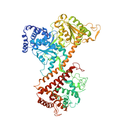
    The SecA ATPase moves polypeptides post-translationally across the plasma membrane of eubacteria, but the mechanism of transport is still unclear. We describe the crystal structure of a novel dimeric form of Bacillus subtilis SecA. Dimerization of SecA occurs at the prominent groove formed by the nucleotide binding domain 2 (nbd2) and the preprotein cross-linking (ppx) domain. The dimer interface is very large, burying approximately 5400 A(2) of solvent accessible surface per monomer. Single cysteine disulfide cross-linking shows the presence of this novel SecA dimer in solution. In addition, other dimers also exist in solution, arguing that they all are in equilibrium with monomeric SecA and supporting the idea that the monomer may be the functional species. Dimerization of SecA causes an alpha-helix of one subunit to convert to a short beta-strand that participates in beta-sheet formation with strands in the other subunit. This conversion of secondary structure elements occurs close to the connection between the nbd1 and ppx domains, a potential site of interaction with translocation substrate. Comparing the different X-ray structures of B. subtilis SecA suggests that small changes in the nucleotide binding domains could be amplified via helix 1 of the helical scaffold domain (hsd) to generate larger movements of the domains involved in polypeptide binding.


  • Organizational Affiliation

    Howard Hughes Medical Institute and Department of Cell Biology, Harvard Medical School, 250 Longwood Avenue, Boston, MA 02115, USA.


Macromolecules
Find similar proteins by:  (by identity cutoff)  |  3D Structure
Entity ID: 1
MoleculeChains Sequence LengthOrganismDetailsImage
Preprotein translocase secA subunit
A, B
780Bacillus subtilisMutation(s): 0 
Gene Names: secAdiv+
UniProt
Find proteins for P28366 (Bacillus subtilis (strain 168))
Explore P28366 
Go to UniProtKB:  P28366
Entity Groups  
Sequence Clusters30% Identity50% Identity70% Identity90% Identity95% Identity100% Identity
UniProt GroupP28366
Sequence Annotations
Expand
  • Reference Sequence
Small Molecules
Ligands 1 Unique
IDChains Name / Formula / InChI Key2D Diagram3D Interactions
ADP
Query on ADP

Download Ideal Coordinates CCD File 
C [auth A]ADENOSINE-5'-DIPHOSPHATE
C10 H15 N5 O10 P2
XTWYTFMLZFPYCI-KQYNXXCUSA-N
Experimental Data & Validation

Experimental Data

  • Method: X-RAY DIFFRACTION
  • Resolution: 3.20 Å
  • R-Value Free: 0.323 
  • R-Value Work: 0.321 
  • R-Value Observed: 0.321 
  • Space Group: I 21 21 21
Unit Cell:
Length ( Å )Angle ( ˚ )
a = 125.834α = 90
b = 166.833β = 90
c = 211.99γ = 90
Software Package:
Software NamePurpose
CNSrefinement
HKL-2000data collection
DENZOdata reduction
SCALEPACKdata scaling
PHASERphasing

Structure Validation

View Full Validation Report


Ligand Structure Quality Assessment 


Entry History 

Deposition Data

Revision History  (Full details and data files)

  • Version 1.0: 2006-11-14
    Type: Initial release
  • Version 1.1: 2008-05-01
    Changes: Version format compliance
  • Version 1.2: 2011-07-13
    Changes: Derived calculations, Version format compliance
  • Version 1.3: 2023-08-30
    Changes: Data collection, Database references, Derived calculations, Refinement description