2HXM

Complex of UNG2 and a small Molecule synthetic Inhibitor


Experimental Data Snapshot

  • Method: X-RAY DIFFRACTION
  • Resolution: 1.30 Å
  • R-Value Free: 0.206 
  • R-Value Work: 0.182 
  • R-Value Observed: 0.183 

wwPDB Validation   3D Report Full Report


Ligand Structure Quality Assessment 


This is version 1.2 of the entry. See complete history


Literature

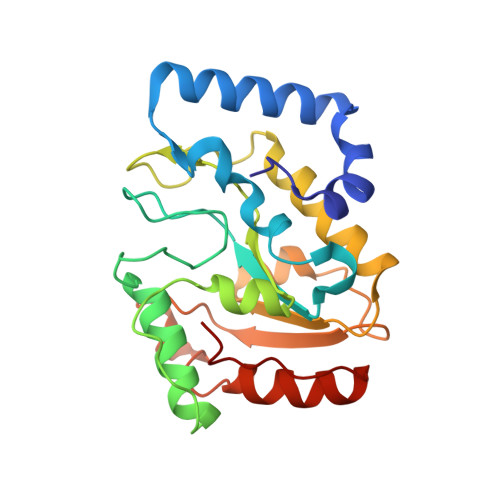
Mimicking damaged DNA with a small molecule inhibitor of human UNG2.

Krosky, D.J.Bianchet, M.A.Seiple, L.Chung, S.Amzel, L.M.Stivers, J.T.

(2006) Nucleic Acids Res 34: 5872-5879

  • DOI: 10.1093/nar/gkl747
  • Primary Citation of Related Structures:  
    2HXM

  • PubMed Abstract: 
  • Human nuclear uracil DNA glycosylase (UNG2) is a cellular DNA repair enzyme that is essential for a number of diverse biological phenomena ranging from antibody diversification to B-cell lymphomas and type-1 human immunodeficiency virus infectivity. During each of these processes, UNG2 recognizes uracilated DNA and excises the uracil base by flipping it into the enzyme active site ...

    Human nuclear uracil DNA glycosylase (UNG2) is a cellular DNA repair enzyme that is essential for a number of diverse biological phenomena ranging from antibody diversification to B-cell lymphomas and type-1 human immunodeficiency virus infectivity. During each of these processes, UNG2 recognizes uracilated DNA and excises the uracil base by flipping it into the enzyme active site. We have taken advantage of the extrahelical uracil recognition mechanism to build large small-molecule libraries in which uracil is tethered via flexible alkane linkers to a collection of secondary binding elements. This high-throughput synthesis and screening approach produced two novel uracil-tethered inhibitors of UNG2, the best of which was crystallized with the enzyme. Remarkably, this inhibitor mimics the crucial hydrogen bonding and electrostatic interactions previously observed in UNG2 complexes with damaged uracilated DNA. Thus, the environment of the binding site selects for library ligands that share these DNA features. This is a general approach to rapid discovery of inhibitors of enzymes that recognize extrahelical damaged bases.


    Related Citations: 
    • Uracil-directed ligand tethering: An efficient strategy for Uracil DNA glycosylase (UNG) inhibitor Development
      Jiang, T.L., Krosky, D.J., Seiple, L., Stivers, J.T.
      (2005) J Am Chem Soc 127: 17412

    Organizational Affiliation

    Department of Pharmacology and Molecular Sciences, 725 North Wolfe Street, Baltimore, MD 21205, USA.


Macromolecules
Find similar proteins by:  (by identity cutoff)  |  3D Structure
Entity ID: 1
MoleculeChainsSequence LengthOrganismDetailsImage
Uracil-DNA glycosylaseA223Homo sapiensMutation(s): 0 
Gene Names: UNGDGUUNG15UNG1
EC: 3.2.2 (PDB Primary Data), 3.2.2.27 (UniProt)
UniProt & NIH Common Fund Data Resources
Find proteins for P13051 (Homo sapiens)
Explore P13051 
Go to UniProtKB:  P13051
PHAROS:  P13051
Entity Groups  
Sequence Clusters30% Identity50% Identity70% Identity90% Identity95% Identity100% Identity
UniProt GroupP13051
Protein Feature View
Expand
  • Reference Sequence
Small Molecules
Ligands 1 Unique
IDChainsName / Formula / InChI Key2D Diagram3D Interactions
302
Query on 302

Download Ideal Coordinates CCD File 
B [auth A]4-[(1E,7E)-8-(2,6-DIOXO-1,2,3,6-TETRAHYDROPYRIMIDIN-4-YL)-3,6-DIOXA-2,7-DIAZAOCTA-1,7-DIEN-1-YL]BENZOIC ACID
C15 H14 N4 O6
XHDKIDMFBWLHAX-GONBZBRSSA-N
 Ligand Interaction
Binding Affinity Annotations 
IDSourceBinding Affinity
302 PDBBind:  2HXM Ki: 6000 (nM) from 1 assay(s)
Experimental Data & Validation

Experimental Data

  • Method: X-RAY DIFFRACTION
  • Resolution: 1.30 Å
  • R-Value Free: 0.206 
  • R-Value Work: 0.182 
  • R-Value Observed: 0.183 
  • Space Group: P 21 21 21
Unit Cell:
Length ( Å )Angle ( ˚ )
a = 43.209α = 90
b = 69.03β = 90
c = 70.318γ = 90
Software Package:
Software NamePurpose
REFMACrefinement
ADSCdata collection
HKL-2000data reduction
SCALEPACKdata scaling
MOLREPphasing

Structure Validation

View Full Validation Report


Ligand Structure Quality Assessment 


Entry History 

Deposition Data

Revision History  (Full details and data files)

  • Version 1.0: 2006-12-05
    Type: Initial release
  • Version 1.1: 2008-05-01
    Changes: Version format compliance
  • Version 1.2: 2011-07-13
    Changes: Version format compliance