2HP0

Crystal structure of iminodisuccinate epimerase


Experimental Data Snapshot

  • Method: X-RAY DIFFRACTION
  • Resolution: 1.50 Å
  • R-Value Free: 0.174 
  • R-Value Work: 0.151 
  • R-Value Observed: 0.152 

wwPDB Validation   3D Report Full Report


Ligand Structure Quality Assessment 


This is version 1.3 of the entry. See complete history


Literature

Three-dimensional Structure of Iminodisuccinate Epimerase Defines the Fold of the MmgE/PrpD Protein Family.

Lohkamp, B.Bauerle, B.Rieger, P.G.Schneider, G.

(2006) J Mol Biol 362: 555-566

  • DOI: https://doi.org/10.1016/j.jmb.2006.07.051
  • Primary Citation of Related Structures:  
    2HP0, 2HP3

  • PubMed Abstract: 

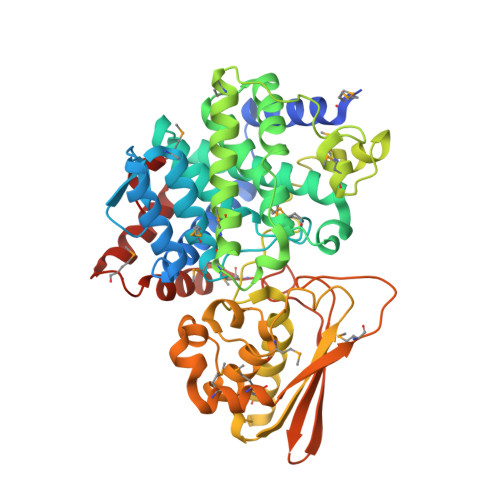
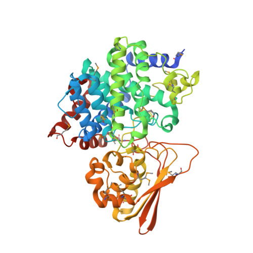
    Iminodisuccinate (IDS) epimerase catalyzes the epimerisation of R,R-, S,S- and R,S- iminodisuccinate, one step in the biodegradation of the chelating agent iminodisuccinate by Agrobacterium tumefaciens BY6. The enzyme is a member of the MmgE/PrpD protein family, a diverse and little characterized class of proteins of prokaryotic and eukaryotic origin. IDS epimerase does not show significant overall amino acid sequence similarity to any other protein of known three-dimensional structure. The crystal structure of this novel epimerase has been determined by multi-wavelength diffraction to 1.5 A resolution using selenomethionine-substituted enzyme. In the crystal, the enzyme forms a homo-dimer, and the subunit consists of two domains. The larger domain, not consecutive in sequence and comprising residues Met1-Lys266 and Leu400-Pro446, forms a novel all alpha-helical fold with a central six-helical bundle. The second, smaller domain folds into an alpha+beta domain, related in topology to chorismate mutase by a circular permutation. IDS epimerase is thus not related in three-dimensional structure to other known epimerases. The fold of the IDS epimerase is representative for the whole MmgE/PrpD family. The putative active site is located at the interface between the two domains of the subunit, and is characterized by a positively charged surface, consistent with the binding of a highly negatively charged substrate such as iminodisuccinate. Docking experiments suggest a two-base mechanism for the epimerisation reaction.


  • Organizational Affiliation

    Department of Medical Biochemistry and Biophysics, Karolinska Institutet, 171 77 Stockholm, Sweden.


Macromolecules
Find similar proteins by:  (by identity cutoff)  |  3D Structure
Entity ID: 1
MoleculeChains Sequence LengthOrganismDetailsImage
IDS-epimerase466Agrobacterium tumefaciensMutation(s): 16 
Gene Names: ite
UniProt
Find proteins for Q1L4E3 (Rhizobium radiobacter)
Explore Q1L4E3 
Go to UniProtKB:  Q1L4E3
Entity Groups  
Sequence Clusters30% Identity50% Identity70% Identity90% Identity95% Identity100% Identity
UniProt GroupQ1L4E3
Sequence Annotations
Expand
  • Reference Sequence
Find similar proteins by:  (by identity cutoff)  |  3D Structure
Entity ID: 2
MoleculeChains Sequence LengthOrganismDetailsImage
IDS-epimerase466Agrobacterium tumefaciensMutation(s): 17 
Gene Names: ite
UniProt
Find proteins for Q1L4E3 (Rhizobium radiobacter)
Explore Q1L4E3 
Go to UniProtKB:  Q1L4E3
Entity Groups  
Sequence Clusters30% Identity50% Identity70% Identity90% Identity95% Identity100% Identity
UniProt GroupQ1L4E3
Sequence Annotations
Expand
  • Reference Sequence
Small Molecules
Ligands 3 Unique
IDChains Name / Formula / InChI Key2D Diagram3D Interactions
DTU
Query on DTU

Download Ideal Coordinates CCD File 
P [auth B],
Q [auth B]
(2R,3S)-1,4-DIMERCAPTOBUTANE-2,3-DIOL
C4 H10 O2 S2
VHJLVAABSRFDPM-ZXZARUISSA-N
EDO
Query on EDO

Download Ideal Coordinates CCD File 
C [auth A]
D [auth A]
E [auth A]
F [auth A]
N [auth B]
C [auth A],
D [auth A],
E [auth A],
F [auth A],
N [auth B],
O [auth B]
1,2-ETHANEDIOL
C2 H6 O2
LYCAIKOWRPUZTN-UHFFFAOYSA-N
UNX
Query on UNX

Download Ideal Coordinates CCD File 
G [auth A]
H [auth A]
I [auth A]
J [auth A]
K [auth A]
G [auth A],
H [auth A],
I [auth A],
J [auth A],
K [auth A],
L [auth A],
M [auth A],
R [auth B],
S [auth B],
T [auth B],
U [auth B],
V [auth B],
W [auth B],
X [auth B]
UNKNOWN ATOM OR ION
X
Modified Residues  2 Unique
IDChains TypeFormula2D DiagramParent
MSE
Query on MSE
A
L-PEPTIDE LINKINGC5 H11 N O2 SeMET
CSO
Query on CSO
B
L-PEPTIDE LINKINGC3 H7 N O3 SCYS
Experimental Data & Validation

Experimental Data

  • Method: X-RAY DIFFRACTION
  • Resolution: 1.50 Å
  • R-Value Free: 0.174 
  • R-Value Work: 0.151 
  • R-Value Observed: 0.152 
  • Space Group: P 1 21 1
Unit Cell:
Length ( Å )Angle ( ˚ )
a = 55.602α = 90
b = 104.973β = 102.51
c = 78.645γ = 90
Software Package:
Software NamePurpose
REFMACrefinement
MOSFLMdata reduction
CCP4data scaling
SOLVEphasing

Structure Validation

View Full Validation Report


Ligand Structure Quality Assessment 


Entry History 

Deposition Data

Revision History  (Full details and data files)

  • Version 1.0: 2006-09-12
    Type: Initial release
  • Version 1.1: 2008-05-01
    Changes: Version format compliance
  • Version 1.2: 2011-07-13
    Changes: Version format compliance
  • Version 1.3: 2011-10-19
    Changes: Derived calculations