2HOS

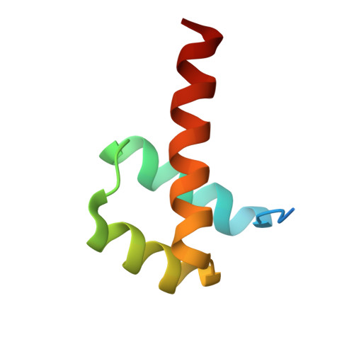
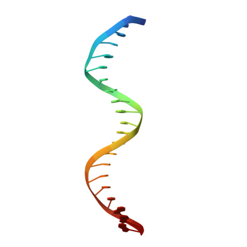
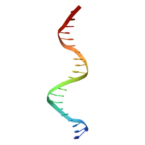
Phage-Selected Homeodomain Bound to Unmodified DNA


Experimental Data Snapshot

  • Method: X-RAY DIFFRACTION
  • Resolution: 1.90 Å
  • R-Value Free: 0.257 
  • R-Value Work: 0.223 
  • R-Value Observed: 0.225 

Starting Model: experimental
View more details

wwPDB Validation   3D Report Full Report


This is version 1.5 of the entry. See complete history


Literature

Structure and properties of a re-engineered homeodomain protein-DNA interface.

Simon, M.D.Feldman, M.E.Rauh, D.Maris, A.E.Wemmer, D.E.Shokat, K.M.

(2006) ACS Chem Biol 1: 755-760

  • DOI: https://doi.org/10.1021/cb6003756
  • Primary Citation of Related Structures:  
    2HOS, 2HOT

  • PubMed Abstract: 

    The homeodomain (HD)-DNA interface has been conserved over 500 million years of evolution. Despite this conservation, we have successfully re-engineered the engrailed HD to specifically recognize an unnatural nucleotide using a phage display selection. Here we report the synthesis of novel nucleosides and the selection of mutant HDs that bind these nucleotides using phage display. The high-resolution crystal structure of one mutant in complex with modified and unmodified DNA demonstrates that, even with the substantial perturbation to the interface, this selected mutant retains a canonical HD structure. Dissection of the contributions due to each of the selected mutations reveals that the majority of the modification-specific binding is accomplished by a single mutation (I47G) but that the remaining mutations retune the stability of the HD. These results afford a detailed look at a re-engineered protein-DNA interaction and provide insight into the opportunities for re-engineering highly conserved interfaces.


Macromolecules

Find similar proteins by:  (by identity cutoff)  |  3D Structure
Entity ID: 3
MoleculeChains Sequence LengthOrganismDetailsImage
Segmentation polarity homeobox protein engrailedC [auth A],
D [auth B]
63Drosophila melanogasterMutation(s): 4 
Gene Names: en
UniProt
Find proteins for P02836 (Drosophila melanogaster)
Explore P02836 
Go to UniProtKB:  P02836
Entity Groups  
Sequence Clusters30% Identity50% Identity70% Identity90% Identity95% Identity100% Identity
UniProt GroupP02836
Sequence Annotations
Expand
  • Reference Sequence

Find similar nucleic acids by:  Sequence   |   3D Structure  

Entity ID: 1
MoleculeChains LengthOrganismImage
5'-D(*TP*TP*TP*TP*GP*CP*CP*AP*TP*GP*TP*AP*AP*TP*CP*CP*CP*CP*GP*GP*A)-3'A [auth C]21N/A
Sequence Annotations
Expand
  • Reference Sequence

Find similar nucleic acids by:  Sequence   |   3D Structure  

Entity ID: 2
MoleculeChains LengthOrganismImage
5'-D(*AP*TP*CP*CP*GP*GP*GP*GP*AP*TP*TP*AP*CP*AP*TP*GP*GP*CP*AP*AP*A)-3'B [auth D]21N/A
Sequence Annotations
Expand
  • Reference Sequence
Experimental Data & Validation

Experimental Data

  • Method: X-RAY DIFFRACTION
  • Resolution: 1.90 Å
  • R-Value Free: 0.257 
  • R-Value Work: 0.223 
  • R-Value Observed: 0.225 
  • Space Group: C 1 2 1
Unit Cell:
Length ( Å )Angle ( ˚ )
a = 127.097α = 90
b = 45.025β = 118.41
c = 73.081γ = 90
Software Package:
Software NamePurpose
DENZOdata reduction
SCALEPACKdata scaling
REFMACrefinement
PDB_EXTRACTdata extraction
HKL-2000data reduction
HKL-2000data scaling
CNSphasing

Structure Validation

View Full Validation Report


Entry History 

Deposition Data

Revision History  (Full details and data files)

  • Version 1.0: 2006-12-12
    Type: Initial release
  • Version 1.1: 2008-05-01
    Changes: Version format compliance
  • Version 1.2: 2011-07-13
    Changes: Advisory, Version format compliance
  • Version 1.3: 2017-10-18
    Changes: Refinement description
  • Version 1.4: 2021-10-20
    Changes: Database references, Derived calculations
  • Version 1.5: 2023-08-30
    Changes: Data collection, Refinement description