


Crystal structure of a singleton protein PF1176 from P. furiosus
Rose, J.P., Liu, Z.-J., Wang, B.-C.To be published.
Experimental Data Snapshot
wwPDB Validation 3D Report Full Report

Entity ID: 1 | |||||
|---|---|---|---|---|---|
| Molecule | Chains | Sequence Length | Organism | Details | Image |
| Hypothetical protein PF1176 | 103 | Pyrococcus furiosus | Mutation(s): 2 Gene Names: pf1176 | 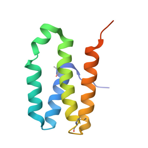 | |
UniProt | |||||
Entity Groups | |||||
| Sequence Clusters | 30% Identity50% Identity70% Identity90% Identity95% Identity100% Identity | ||||
| UniProt Group | Q8U1N0 | ||||
Sequence AnnotationsExpand | |||||
Reference Sequence | |||||
| Modified Residues 1 Unique | |||||
|---|---|---|---|---|---|
| ID | Chains | Type | Formula | 2D Diagram | Parent |
| MSE Query on MSE | A, B, C, D | L-PEPTIDE LINKING | C5 H11 N O2 Se |  | MET |
| Length ( Å ) | Angle ( ˚ ) |
|---|---|
| a = 62.68 | α = 90 |
| b = 64.24 | β = 90 |
| c = 111.65 | γ = 90 |
| Software Name | Purpose |
|---|---|
| SCA2STRUCTURE | model building |
| CNS | refinement |
| HKL-2000 | data reduction |
| HKL-2000 | data scaling |
| SCA2STRUCTURE | phasing |


