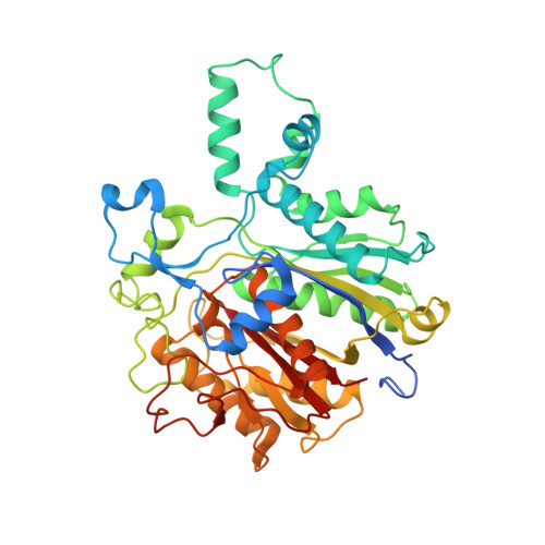
X-Ray Crystal Structure of Mycobacterium tuberculosis beta-Ketoacyl Acyl Carrier Protein Synthase II (mtKasB).
Sridharan, S., Wang, L., Brown, A.K., Dover, L.G., Kremer, L., Besra, G.S., Sacchettini, J.C.(2007) J Mol Biology 366: 469-480
- PubMed: 17174327 Search on PubMedSearch on PubMed Central
- DOI: https://doi.org/10.1016/j.jmb.2006.11.006
- Primary Citation Related Structures:
2GP6 - PubMed Abstract:
Mycolic acids are long chain alpha-alkyl branched, beta-hydroxy fatty acids that represent a characteristic component of the Mycobacterium tuberculosis cell wall. Through their covalent attachment to peptidoglycan via an arabinogalactan polysaccharide, they provide the basis for an essential outer envelope membrane. Mycobacteria possess two fatty acid synthases (FAS); FAS-I carries out de novo synthesis of fatty acids while FAS-II is considered to elongate medium chain length fatty acyl primers to provide long chain (C(56)) precursors of mycolic acids. Here we report the crystal structure of Mycobacterium tuberculosis beta-ketoacyl acyl carrier protein synthase (ACP) II mtKasB, a mycobacterial elongation condensing enzyme involved in FAS-II. This enzyme, along with the M. tuberculosis beta-ketoacyl ACP synthase I mtKasA, catalyzes the Claisen-type condensation reaction responsible for fatty acyl elongation in FAS-II and are potential targets for development of novel anti-tubercular drugs. The crystal structure refined to 2.4 A resolution revealed that, like other KAS-II enzymes, mtKasB adopts a thiolase fold but contains unique structural features in the capping region that may be crucial to its preference for longer fatty acyl chains than its counterparts from other bacteria. Modeling of mtKasA using the mtKasB structure as a template predicts the overall structures to be almost identical, but a larger entrance to the active site tunnel is envisaged that might contribute to the greater sensitivity of mtKasA to the inhibitor thiolactomycin (TLM). Modeling of TLM binding in mtKasB shows that the drug fits the active site poorly and results of enzyme inhibition assays using TLM analogues are wholly consistent with our structural observations. Consequently, the structure described here further highlights the potential of TLM as an anti-tubercular lead compound and will aid further exploration of the TLM scaffold towards the design of novel compounds, which inhibit mycobacterial KAS enzymes more effectively.
- Department of Biochemistry and Biophysics, Texas A&M University, College Station, TX 77843-2128, USA.
Organizational Affiliation: