2G9J

Complex of TM1a(1-14)Zip with TM9a(251-284): a model for the polymerization domain ("overlap region") of tropomyosin, Northeast Structural Genomics Target OR9


Experimental Data Snapshot

  • Method: SOLUTION NMR
  • Conformers Calculated: 196 
  • Conformers Submitted: 10 
  • Selection Criteria: 10 structures from initial DYANA calcultions with the lowest target functions were refined using CNS. The structures back calculated data agree with experimental NOESY spectra. The structures have acceptable covalent geometry, favorable non-bond energy, the lowest energy and the fewest restraint violations. 

wwPDB Validation   3D Report Full Report


This is version 1.3 of the entry. See complete history


Literature

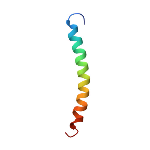
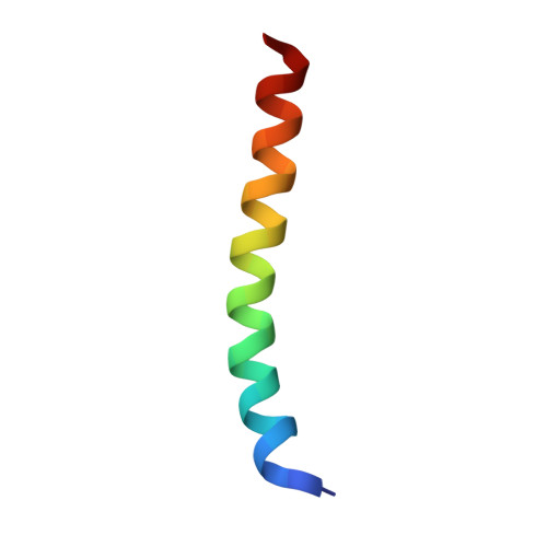
Solution NMR Structure of the Junction between Tropomyosin Molecules: Implications for Actin Binding and Regulation.

Greenfield, N.J.Huang, Y.J.Swapna, G.V.Bhattacharya, A.Rapp, B.Singh, A.Montelione, G.T.Hitchcock-Degregori, S.E.

(2006) J Mol Biol 364: 80-96

  • DOI: https://doi.org/10.1016/j.jmb.2006.08.033
  • Primary Citation of Related Structures:  
    2G9J

  • PubMed Abstract: 

    Tropomyosin is a coiled-coil protein that binds head-to-tail along the length of actin filaments in eukaryotic cells, stabilizing them and providing protection from severing proteins. Tropomyosin cooperatively regulates actin's interaction with myosin and mediates the Ca2+ -dependent regulation of contraction by troponin in striated muscles. The N-terminal and C-terminal ends are critical functional determinants that form an "overlap complex". Here we report the solution NMR structure of an overlap complex formed of model peptides. In the complex, the chains of the C-terminal coiled coil spread apart to allow insertion of 11 residues of the N-terminal coiled coil into the resulting cleft. The plane of the N-terminal coiled coil is rotated 90 degrees relative to the plane of the C terminus. A consequence of the geometry is that the orientation of postulated periodic actin binding sites on the coiled-coil surface is retained from one molecule to the next along the actin filament when the overlap complex is modeled into the X-ray structure of tropomyosin determined at 7 Angstroms. Nuclear relaxation NMR data reveal flexibility of the junction, which may function to optimize binding along the helical actin filament and to allow mobility of tropomyosin on the filament surface as it switches between regulatory states.


  • Organizational Affiliation

    Department of Neuroscience and Cell Biology, Robert Wood Johnson Medical School, Piscataway, NJ 08854, USA. greenfie@umdnj.edu


Macromolecules
Find similar proteins by:  (by identity cutoff)  |  3D Structure
Entity ID: 1
MoleculeChains Sequence LengthOrganismDetailsImage
Tropomyosin 1 alpha chainA [auth C],
C [auth D]
37Rattus norvegicusMutation(s): 1 
Gene Names: TPM1
UniProt
Find proteins for P04692 (Rattus norvegicus)
Explore P04692 
Go to UniProtKB:  P04692
Entity Groups  
Sequence Clusters30% Identity50% Identity70% Identity90% Identity95% Identity100% Identity
UniProt GroupP04692
Sequence Annotations
Expand
  • Reference Sequence
Find similar proteins by:  (by identity cutoff)  |  3D Structure
Entity ID: 2
MoleculeChains Sequence LengthOrganismDetailsImage
Tropomyosin 1 alpha chain/General control protein GCN4B [auth A],
D [auth B]
33Rattus norvegicusSaccharomyces cerevisiae
This entity is chimeric
Mutation(s): 0 
Gene Names: TPM1
UniProt
Find proteins for P03069 (Saccharomyces cerevisiae (strain ATCC 204508 / S288c))
Explore P03069 
Go to UniProtKB:  P03069
Find proteins for P04692 (Rattus norvegicus)
Explore P04692 
Go to UniProtKB:  P04692
Entity Groups  
Sequence Clusters30% Identity50% Identity70% Identity90% Identity95% Identity100% Identity
UniProt GroupsP03069P04692
Sequence Annotations
Expand
  • Reference Sequence
Experimental Data & Validation

Experimental Data

  • Method: SOLUTION NMR
  • Conformers Calculated: 196 
  • Conformers Submitted: 10 
  • Selection Criteria: 10 structures from initial DYANA calcultions with the lowest target functions were refined using CNS. The structures back calculated data agree with experimental NOESY spectra. The structures have acceptable covalent geometry, favorable non-bond energy, the lowest energy and the fewest restraint violations. 

Structure Validation

View Full Validation Report


Entry History 

Revision History  (Full details and data files)

  • Version 1.0: 2006-11-07
    Type: Initial release
  • Version 1.1: 2008-05-01
    Changes: Version format compliance
  • Version 1.2: 2011-07-13
    Changes: Version format compliance
  • Version 1.3: 2021-10-20
    Changes: Data collection, Database references, Derived calculations