Active-site changes in the pyruvate dehydrogenase multienzyme complex E1 apoenzyme component from Escherichia coli observed at 2.32 A resolution.
Chandrasekhar, K., Arjunan, P., Sax, M., Nemeria, N., Jordan, F., Furey, W.(2006) Acta Crystallogr D Biol Crystallogr 62: 1382-1386
- PubMed: 17057342 Search on PubMed
- DOI: https://doi.org/10.1107/S0907444906034408
- Primary Citation Related Structures:
2G67 - PubMed Abstract:
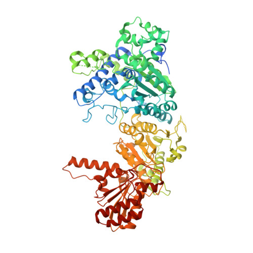
The first enzymatic component, E1 (EC 1.2.4.1), of the pyruvate dehydrogenase multienzyme complex (PDHc) utilizes thiamine diphosphate (ThDP) and Mg(2+) as cofactors. The structure of a branched-chain-specific E1 apoenzyme from the heterotetrameric alpha(2)beta(2) E1 family was recently reported and showed that disorder-to-order transformations in two active-site loops take place upon cofactor binding. To ascertain what effect the absence of cofactor may have in the homodimeric alpha(2) Escherichia coli PDHc E1, the corresponding apoenzyme has been prepared and its three-dimensional structure determined and analyzed at 2.32 A by crystallographic methods. This represents the first reported apoenzyme structure for any E1 component from the homodimeric alpha(2) family. Electron-density features occurring in the region where the cofactor pyrimidine ring would normally be expected to bind are of size, shape and location compatible with water molecules that form a hydrogen-bonded linkage between residues Glu571 and Val192, which normally make conserved interactions with the ThDP cofactor. A histidine side chain that normally forms hydrogen bonds to ThDP is disordered in its absence and partially occupies two sites. Unlike in the reported heterotetrameric branched-chain apo-E1, no disorder/order loop transformations are evident in apo-PDHc E1 relative to the holo-E1 enzyme (PDHc E1-ThDP-Mg(2+)). Differences in the extent of hydrogen-bonding networks found in the apo-E1 enzyme, the holo-E1 enzyme and in an inhibitor complex with bound thiamine 2-thiazolone diphosphate (ThTDP), PDHc E1-ThTDP-Mg(2+), are described.
- Biocrystallography Laboratory, Veterans Affairs Medical Center, PO Box 12055, University Drive C, Pittsburgh, PA 15240, USA.
Organizational Affiliation: