2G4B

Structure of U2AF65 variant with polyuridine tract


Experimental Data Snapshot

  • Method: X-RAY DIFFRACTION
  • Resolution: 2.50 Å
  • R-Value Free: 0.276 
  • R-Value Work: 0.261 
  • R-Value Observed: 0.262 

wwPDB Validation   3D Report Full Report


This is version 1.5 of the entry. See complete history


Literature

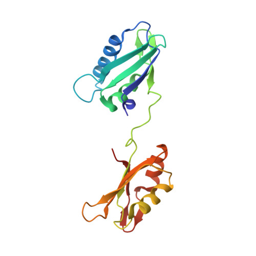
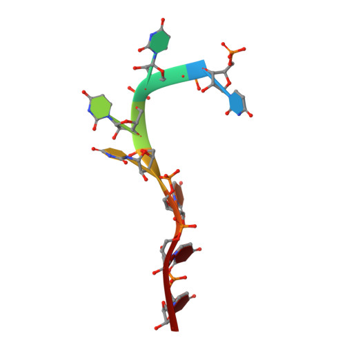
Structural basis of polypyrimidine tract recognition by the essential splicing factor U2AF65.

Sickmier, E.A.Frato, K.E.Paranawithana, S.Shen, H.Green, M.R.Kielkopf, C.L.

(2006) Mol Cell 23: 49-59

  • DOI: https://doi.org/10.1016/j.molcel.2006.05.025
  • Primary Citation of Related Structures:  
    2G4B

  • PubMed Abstract: 

    The essential pre-mRNA splicing factor, U2AF(65), guides the early stages of splice site choice by recognizing a polypyrimidine (Py) tract consensus sequence near the 3' splice site. Since Py tracts are relatively poorly conserved in higher eukaryotes, U2AF(65) is faced with the problem of specifying uridine-rich sequences, yet tolerating a variety of nucleotide substitutions found in natural Py tracts. To better understand these apparently contradictory RNA binding characteristics, the X-ray structure of the U2AF(65) RNA binding domain bound to a Py tract composed of seven uridines has been determined at 2.5 A resolution. Specific hydrogen bonds between U2AF(65) and the uracil bases provide an explanation for polyuridine recognition. Flexible side chains and bound water molecules form the majority of the base contacts and potentially could rearrange when the U2AF(65) structure adapts to different Py tract sequences. The energetic importance of conserved residues for Py tract binding is established by analysis of site-directed mutant U2AF(65) proteins using surface plasmon resonance.


  • Organizational Affiliation

    Department of Biochemistry and Molecular Biology, Johns Hopkins University Bloomberg School of Public Health, Baltimore, Maryland 21205, USA.


Macromolecules

Find similar proteins by:  (by identity cutoff)  |  3D Structure
Entity ID: 2
MoleculeChains Sequence LengthOrganismDetailsImage
Splicing factor U2AF 65 kDa subunitB [auth A]172Homo sapiensMutation(s): 0 
Gene Names: U2AF65
UniProt & NIH Common Fund Data Resources
Find proteins for P26368 (Homo sapiens)
Explore P26368 
Go to UniProtKB:  P26368
PHAROS:  P26368
GTEx:  ENSG00000063244 
Entity Groups  
Sequence Clusters30% Identity50% Identity70% Identity90% Identity95% Identity100% Identity
UniProt GroupP26368
Sequence Annotations
Expand
  • Reference Sequence

Find similar nucleic acids by:  Sequence   |   3D Structure  

Entity ID: 1
MoleculeChains LengthOrganismImage
5'-R(P*UP*UP*UP*UP*UP*UP*U)-3'A [auth B]7N/A
Sequence Annotations
Expand
  • Reference Sequence
Small Molecules
Ligands 1 Unique
IDChains Name / Formula / InChI Key2D Diagram3D Interactions
DIO
Query on DIO

Download Ideal Coordinates CCD File 
C [auth A]1,4-DIETHYLENE DIOXIDE
C4 H8 O2
RYHBNJHYFVUHQT-UHFFFAOYSA-N
Binding Affinity Annotations 
IDSourceBinding Affinity
RNA PDBBind:  2G4B Kd: 1800 (nM) from 1 assay(s)
Experimental Data & Validation

Experimental Data

  • Method: X-RAY DIFFRACTION
  • Resolution: 2.50 Å
  • R-Value Free: 0.276 
  • R-Value Work: 0.261 
  • R-Value Observed: 0.262 
  • Space Group: P 65 2 2
Unit Cell:
Length ( Å )Angle ( ˚ )
a = 129.388α = 90
b = 129.388β = 90
c = 62.645γ = 120
Software Package:
Software NamePurpose
HKL-2000data collection
SCALEPACKdata scaling
CNSrefinement
HKL-2000data reduction
CNSphasing

Structure Validation

View Full Validation Report


Entry History 

Deposition Data

Revision History  (Full details and data files)

  • Version 1.0: 2006-07-25
    Type: Initial release
  • Version 1.1: 2008-05-01
    Changes: Version format compliance
  • Version 1.2: 2011-07-13
    Changes: Version format compliance
  • Version 1.3: 2017-08-16
    Changes: Refinement description, Source and taxonomy
  • Version 1.4: 2018-01-31
    Changes: Database references
  • Version 1.5: 2023-08-30
    Changes: Data collection, Database references, Derived calculations, Refinement description