2FWO

MHC Class I H-2Kd heavy chain in complex with beta-2microglobulin and peptide derived from influenza nucleoprotein


Experimental Data Snapshot

  • Method: X-RAY DIFFRACTION
  • Resolution: 2.60 Å
  • R-Value Free: 0.269 
  • R-Value Work: 0.218 
  • R-Value Observed: 0.252 

wwPDB Validation   3D Report Full Report


This is version 1.4 of the entry. See complete history


Literature

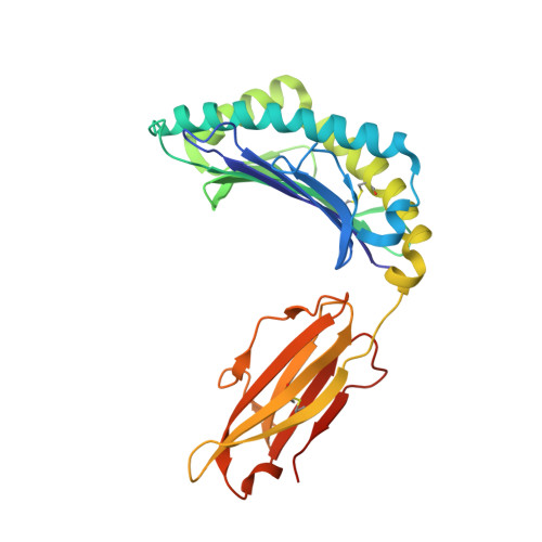
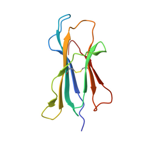
Structural Definition of the H-2Kd Peptide-binding Motif.

Mitaksov, V.Fremont, D.H.

(2006) J Biol Chem 281: 10618-10625

  • DOI: https://doi.org/10.1074/jbc.M510511200
  • Primary Citation of Related Structures:  
    2FWO

  • PubMed Abstract: 

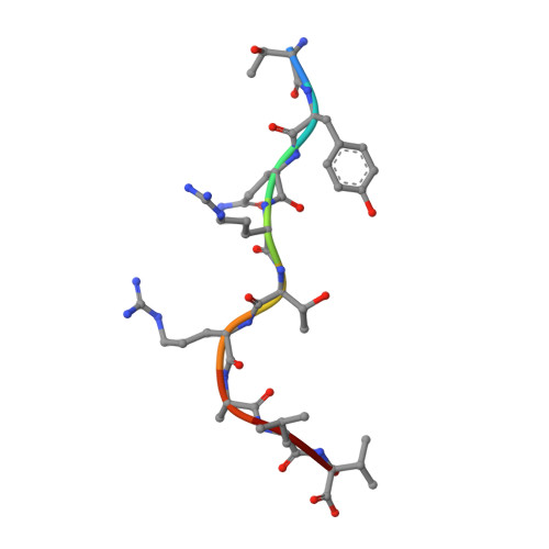
    Classic major histocompatibility complex (MHC) proteins associate with antigen- and self-derived peptides in an allele-specific manner. Herein we present the crystal structure of the MHC class I protein H-2K(d) (K(d)) expressed by BALB/c mice in complex with an antigenic peptide derived from influenza A/PR/8/34 nucleoprotein (Flu, residues 147-155, TYQRTRALV). Analysis of our structure in conjunction with the sequences of naturally processed epitopes provides a comprehensive understanding of the dominant K(d) peptide-binding motif. We find that Flu residues Tyr(P2), Thr(P5), and Val(P9) are sequestered into the B, C, and F pockets of the K(d) groove, respectively. The shape and chemistry of the polymorphic B pocket make it an optimal binding site for the side chain of Tyr(P2) as the dominant anchoring residue of nonameric peptides. The non-polar F pocket limits the amino acid repertoire at P9 to hydrophobic residues such as Ile, Leu, or Val, whereas the C pocket restricts the size of the P5-anchoring side chain. We also show that Flu is accommodated in the complex through an unfavorable kink in the otherwise extended peptide backbone due to the presence of a prominent ridge in the K(d) groove. Surprisingly, this backbone conformation is strikingly similar to D(b)-presented peptides despite the fact that these proteins employ distinct motif-anchoring strategies. The results presented in this study provide a solid foundation for the understanding of K(d)-restricted antigen presentation and recognition events.


  • Organizational Affiliation

    Department of Pathology and Immunology, Washington University School of Medicine, 660 South Euclid Avenue, St. Louis, MO 63110, USA.


Macromolecules
Find similar proteins by:  (by identity cutoff)  |  3D Structure
Entity ID: 1
MoleculeChains Sequence LengthOrganismDetailsImage
H-2 class I histocompatibility antigen, K-D alpha chain283Mus musculusMutation(s): 0 
Gene Names: H2-K1H2-K
UniProt
Find proteins for P01902 (Mus musculus)
Explore P01902 
Go to UniProtKB:  P01902
Entity Groups  
Sequence Clusters30% Identity50% Identity70% Identity90% Identity95% Identity100% Identity
UniProt GroupP01902
Sequence Annotations
Expand
  • Reference Sequence
Find similar proteins by:  (by identity cutoff)  |  3D Structure
Entity ID: 2
MoleculeChains Sequence LengthOrganismDetailsImage
Beta-2-microglobulin100Mus musculusMutation(s): 0 
Gene Names: B2m
UniProt & NIH Common Fund Data Resources
Find proteins for P01887 (Mus musculus)
Explore P01887 
Go to UniProtKB:  P01887
IMPC:  MGI:88127
Entity Groups  
Sequence Clusters30% Identity50% Identity70% Identity90% Identity95% Identity100% Identity
UniProt GroupP01887
Sequence Annotations
Expand
  • Reference Sequence

Find similar proteins by:  Sequence   |   3D Structure  

Entity ID: 3
MoleculeChains Sequence LengthOrganismDetailsImage
TYQRTRALV peptide from NucleoproteinC [auth P]9N/AMutation(s): 0 
UniProt
Find proteins for Q701N7 (Influenza A virus)
Explore Q701N7 
Go to UniProtKB:  Q701N7
Entity Groups  
UniProt GroupQ701N7
Sequence Annotations
Expand
  • Reference Sequence
Experimental Data & Validation

Experimental Data

  • Method: X-RAY DIFFRACTION
  • Resolution: 2.60 Å
  • R-Value Free: 0.269 
  • R-Value Work: 0.218 
  • R-Value Observed: 0.252 
  • Space Group: P 21 21 21
Unit Cell:
Length ( Å )Angle ( ˚ )
a = 117.2α = 90
b = 85.3β = 90
c = 42.6γ = 90
Software Package:
Software NamePurpose
DENZOdata reduction
SCALEPACKdata scaling
AMoREphasing
CNSrefinement
PDB_EXTRACTdata extraction

Structure Validation

View Full Validation Report


Entry History 

Deposition Data

Revision History  (Full details and data files)

  • Version 1.0: 2006-02-21
    Type: Initial release
  • Version 1.1: 2008-05-01
    Changes: Version format compliance
  • Version 1.2: 2011-07-13
    Changes: Version format compliance
  • Version 1.3: 2017-10-18
    Changes: Refinement description
  • Version 1.4: 2023-08-30
    Changes: Data collection, Database references, Refinement description