2FPR

Crystal structure the N-terminal domain of E. coli HisB. Apo Mg model.


Experimental Data Snapshot

  • Method: X-RAY DIFFRACTION
  • Resolution: 1.70 Å
  • R-Value Free: 0.220 
  • R-Value Work: 0.189 
  • R-Value Observed: 0.190 

wwPDB Validation   3D Report Full Report


This is version 1.4 of the entry. See complete history


Literature

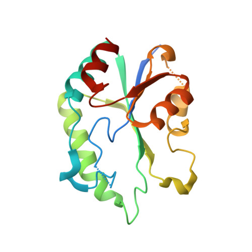
Structural snapshots of Escherichia coli histidinol phosphate phosphatase along the reaction pathway.

Rangarajan, E.S.Proteau, A.Wagner, J.Hung, M.N.Matte, A.Cygler, M.

(2006) J Biol Chem 281: 37930-37941

  • DOI: https://doi.org/10.1074/jbc.M604916200
  • Primary Citation of Related Structures:  
    2FPR, 2FPS, 2FPU, 2FPW, 2FPX

  • PubMed Abstract: 

    HisB from Escherichia coli is a bifunctional enzyme catalyzing the sixth and eighth steps of l-histidine biosynthesis. The N-terminal domain (HisB-N) possesses histidinol phosphate phosphatase activity, and its crystal structure shows a single domain with fold similarity to the haloacid dehalogenase (HAD) enzyme family. HisB-N forms dimers in the crystal and in solution. The structure shows the presence of a structural Zn(2+) ion stabilizing the conformation of an extended loop. Two metal binding sites were also identified in the active site. Their presence was further confirmed by isothermal titration calorimetry. HisB-N is active in the presence of Mg(2+), Mn(2+), Co(2+), or Zn(2+), but Ca(2+) has an inhibitory effect. We have determined structures of several intermediate states corresponding to snapshots along the reaction pathway, including that of the phosphoaspartate intermediate. A catalytic mechanism, different from that described for other HAD enzymes, is proposed requiring the presence of the second metal ion not found in the active sites of previously characterized HAD enzymes, to complete the second half-reaction. The proposed mechanism is reminiscent of two-Mg(2+) ion catalysis utilized by DNA and RNA polymerases and many nucleases. The structure also provides an explanation for the inhibitory effect of Ca(2+).


  • Organizational Affiliation

    Department of Biochemistry, McGill University, Montréal, Québec H3G 1Y6, Canada.


Macromolecules
Find similar proteins by:  (by identity cutoff)  |  3D Structure
Entity ID: 1
MoleculeChains Sequence LengthOrganismDetailsImage
Histidine biosynthesis bifunctional protein hisB
A, B
176Escherichia coli O157:H7Mutation(s): 0 
Gene Names: hisB
EC: 3.1.3.15
UniProt
Find proteins for Q9S5G5 (Escherichia coli O157:H7)
Explore Q9S5G5 
Go to UniProtKB:  Q9S5G5
Entity Groups  
Sequence Clusters30% Identity50% Identity70% Identity90% Identity95% Identity100% Identity
UniProt GroupQ9S5G5
Sequence Annotations
Expand
  • Reference Sequence
Small Molecules
Ligands 4 Unique
IDChains Name / Formula / InChI Key2D Diagram3D Interactions
BR
Query on BR

Download Ideal Coordinates CCD File 
E [auth A]
F [auth A]
G [auth A]
K [auth B]
L [auth B]
E [auth A],
F [auth A],
G [auth A],
K [auth B],
L [auth B],
M [auth B],
N [auth B]
BROMIDE ION
Br
CPELXLSAUQHCOX-UHFFFAOYSA-M
ZN
Query on ZN

Download Ideal Coordinates CCD File 
C [auth A],
H [auth B]
ZINC ION
Zn
PTFCDOFLOPIGGS-UHFFFAOYSA-N
MG
Query on MG

Download Ideal Coordinates CCD File 
D [auth A],
I [auth B]
MAGNESIUM ION
Mg
JLVVSXFLKOJNIY-UHFFFAOYSA-N
NA
Query on NA

Download Ideal Coordinates CCD File 
J [auth B]SODIUM ION
Na
FKNQFGJONOIPTF-UHFFFAOYSA-N
Experimental Data & Validation

Experimental Data

  • Method: X-RAY DIFFRACTION
  • Resolution: 1.70 Å
  • R-Value Free: 0.220 
  • R-Value Work: 0.189 
  • R-Value Observed: 0.190 
  • Space Group: C 2 2 21
Unit Cell:
Length ( Å )Angle ( ˚ )
a = 53.256α = 90
b = 131.962β = 90
c = 106.14γ = 90
Software Package:
Software NamePurpose
REFMACrefinement
HKL-2000data reduction
SCALEPACKdata scaling
SHELXDphasing

Structure Validation

View Full Validation Report


Entry History 

Deposition Data

Revision History  (Full details and data files)

  • Version 1.0: 2006-09-05
    Type: Initial release
  • Version 1.1: 2008-05-01
    Changes: Version format compliance
  • Version 1.2: 2011-07-13
    Changes: Source and taxonomy, Version format compliance
  • Version 1.3: 2018-01-24
    Changes: Structure summary
  • Version 1.4: 2024-02-14
    Changes: Data collection, Database references, Derived calculations