2FOJ

The Crystal Structure of the N-terminal domain of HAUSP/USP7 complexed with p53 peptide 364-367


Experimental Data Snapshot

  • Method: X-RAY DIFFRACTION
  • Resolution: 1.60 Å
  • R-Value Free: 0.262 
  • R-Value Work: 0.229 
  • R-Value Observed: 0.229 

wwPDB Validation   3D Report Full Report


This is version 1.4 of the entry. See complete history


Literature

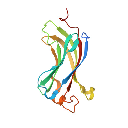
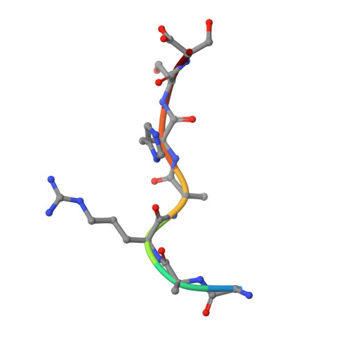
Molecular recognition of p53 and MDM2 by USP7/HAUSP

Sheng, Y.Saridakis, V.Sarkari, F.Duan, S.Wu, T.Arrowsmith, C.H.Frappier, L.

(2006) Nat Struct Mol Biol 13: 285-291

  • DOI: https://doi.org/10.1038/nsmb1067
  • Primary Citation of Related Structures:  
    2FOJ, 2FOO, 2FOP

  • PubMed Abstract: 
  • The ubiquitin-specific protease, USP7, has key roles in the p53 pathway whereby it stabilizes both p53 and MDM2. We show that the N-terminal domain of USP7 binds two closely spaced 4-residue sites in both p53 and MDM2, falling between p53 residues 359-367 and MDM2 residues 147-159 ...

    The ubiquitin-specific protease, USP7, has key roles in the p53 pathway whereby it stabilizes both p53 and MDM2. We show that the N-terminal domain of USP7 binds two closely spaced 4-residue sites in both p53 and MDM2, falling between p53 residues 359-367 and MDM2 residues 147-159. Cocrystal structures with USP7 were determined for both p53 peptides and for one MDM2 peptide. These peptides bind the same surface of USP7 as Epstein-Barr nuclear antigen-1, explaining the competitive nature of the interactions. The structures and mutagenesis data indicate a preference for a P/AXXS motif in peptides that bind USP7. Contacts made by serine are identical and crucial for all peptides, and Trp165 in the peptide-binding pocket of USP7 is also crucial. These results help to elucidate the mechanism of substrate recognition by USP7 and the regulation of the p53 pathway.


    Organizational Affiliation

    Ontario Cancer Institute, Department of Medical Biophysics, 101 College Street, Toronto, Ontario, Canada M5G 1L7.


Macromolecules
Find similar proteins by:  (by identity cutoff)  |  3D Structure
Entity ID: 1
MoleculeChainsSequence LengthOrganismDetailsImage
Ubiquitin carboxyl-terminal hydrolase 7155Homo sapiensMutation(s): 0 
Gene Names: USP7HAUSP
EC: 3.1.2.15 (PDB Primary Data), 3.4.19.12 (UniProt)
UniProt & NIH Common Fund Data Resources
Find proteins for Q93009 (Homo sapiens)
Explore Q93009 
Go to UniProtKB:  Q93009
PHAROS:  Q93009
Entity Groups  
Sequence Clusters30% Identity50% Identity70% Identity90% Identity95% Identity100% Identity
UniProt GroupQ93009
Protein Feature View
Expand
  • Reference Sequence

Find similar proteins by:  Sequence   |   3D Structure  

Entity ID: 2
MoleculeChainsSequence LengthOrganismDetailsImage
p53 peptide7N/AMutation(s): 0 
UniProt & NIH Common Fund Data Resources
Find proteins for P04637 (Homo sapiens)
Explore P04637 
Go to UniProtKB:  P04637
PHAROS:  P04637
Entity Groups  
UniProt GroupP04637
Protein Feature View
Expand
  • Reference Sequence
Experimental Data & Validation

Experimental Data

  • Method: X-RAY DIFFRACTION
  • Resolution: 1.60 Å
  • R-Value Free: 0.262 
  • R-Value Work: 0.229 
  • R-Value Observed: 0.229 
  • Space Group: P 41
Unit Cell:
Length ( Å )Angle ( ˚ )
a = 70α = 90
b = 70β = 90
c = 45.6γ = 90
Software Package:
Software NamePurpose
CNSrefinement
HKL-2000data reduction
SCALEPACKdata scaling
CNSphasing

Structure Validation

View Full Validation Report


Entry History 

Deposition Data

Revision History  (Full details and data files)

  • Version 1.0: 2006-02-14
    Type: Initial release
  • Version 1.1: 2008-05-01
    Changes: Version format compliance
  • Version 1.2: 2011-07-13
    Changes: Version format compliance
  • Version 1.3: 2016-12-21
    Changes: Structure summary
  • Version 1.4: 2023-08-30
    Changes: Data collection, Database references, Refinement description