2FJR | pdb_00002fjr

Crystal Structure of Bacteriophage 186


Experimental Data Snapshot

  • Method: X-RAY DIFFRACTION
  • Resolution: 1.95 Å
  • R-Value Free: 
    0.247 (Depositor) 
  • R-Value Work: 
    0.218 (Depositor) 
  • R-Value Observed: 
    0.218 (Depositor) 

wwPDB Validation 3D Report Full Report

Validation slider image for 2FJR

This is version 1.4 of the entry. See complete history

Literature

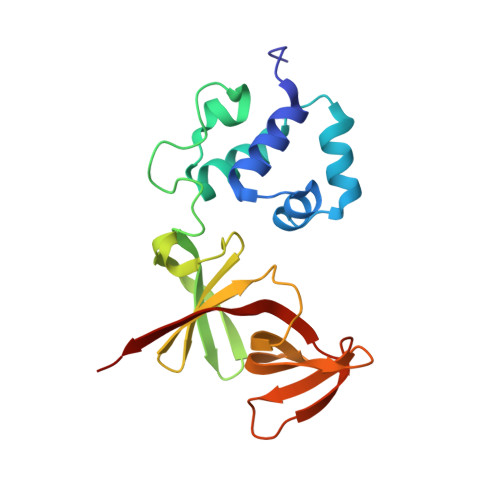
The structural basis of cooperative regulation at an alternate genetic switch

Pinkett, H.Shearwin, K.E.Stayrook, S.Dodd, I.B.Burr, T.Hochschild, A.Egan, J.B.Lewis, M.

(2006) Mol Cell 21: 605-615

  • DOI: https://doi.org/10.1016/j.molcel.2006.01.019
  • Primary Citation Related Structures: 
    2FJR, 2FKD

  • PubMed Abstract: 

    Bacteriophage lambda is a paradigm for understanding the role of cooperativity in gene regulation. Comparison of the regulatory regions of lambda and the unrelated temperate bacteriophage 186 provides insight into alternate ways to assemble functional genetic switches. The structure of the C-terminal domain of the 186 repressor, determined at 2.7 A resolution, reveals an unusual heptamer of dimers, consistent with presented genetic studies. In addition, the structure of a cooperativity mutant of the full-length 186 repressor, identified by genetic screens, was solved to 1.95 A resolution. These structures provide a molecular basis for understanding lysogenic regulation in 186. Whereas the overall fold of the 186 and lambda repressor monomers is remarkably similar, the way the two repressors cooperatively assemble is quite different and explains in part the differences in their regulatory activity.


  • Organizational Affiliation
    • Department of Biochemistry and Biophysics, University of Pennsylvania School of Medicine, 37th and Hamilton Walk, Philadelphia, 19102, USA.

Macromolecule Content 

  • Total Structure Weight: 41.56 kDa 
  • Atom Count: 3,224 
  • Modeled Residue Count: 378 
  • Deposited Residue Count: 378 
  • Unique protein chains: 1

Macromolecules

Find similar proteins by:|  3D Structure
Entity ID: 1
MoleculeChains  Sequence LengthOrganismDetailsImage
Repressor protein CI
A, B
189Eganvirus ev186Mutation(s): 1 
Gene Names: CI
UniProt
Find proteins for P08707 (Escherichia phage 186)
Explore P08707 
Go to UniProtKB:  P08707
Entity Groups
Sequence Clusters30% Identity50% Identity70% Identity90% Identity95% Identity100% Identity
UniProt GroupP08707
Sequence Annotations
Expand
Reference Sequence

Experimental Data & Validation

Experimental Data

  • Method: X-RAY DIFFRACTION
  • Resolution: 1.95 Å
  • R-Value Free:  0.247 (Depositor) 
  • R-Value Work:  0.218 (Depositor) 
  • R-Value Observed: 0.218 (Depositor) 
Space Group: P 41 21 2
Unit Cell:
Length ( Å )Angle ( ˚ )
a = 109.137α = 90
b = 109.137β = 90
c = 93.092γ = 90
Software Package:
Software NamePurpose
CNSrefinement
DENZOdata reduction
SCALEPACKdata scaling
AMoREphasing

Structure Validation

View Full Validation Report


Entry History 

Deposition Data

  • Released Date: 2006-06-13 
  • Deposition Author(s): Lewis, M.

Revision History  (Full details and data files)

  • Version 1.0: 2006-06-13
    Type: Initial release
  • Version 1.1: 2008-05-01
    Changes: Version format compliance
  • Version 1.2: 2011-07-13
    Changes: Version format compliance
  • Version 1.3: 2021-10-20
    Changes: Database references
  • Version 1.4: 2024-02-14
    Changes: Data collection