Solution structure of p53 core domain: Structural basis for its instability
Perez-Canadillas, J.M., Tidow, H., Freund, S.M., Rutherford, T.J., Ang, H.C., Fersht, A.R.(2006) Proc Natl Acad Sci U S A 103: 2109-2114
- PubMed: 16461916 Search on PubMedSearch on PubMed Central
- DOI: https://doi.org/10.1073/pnas.0510941103
- Primary Citation Related Structures:
2FEJ - PubMed Abstract:
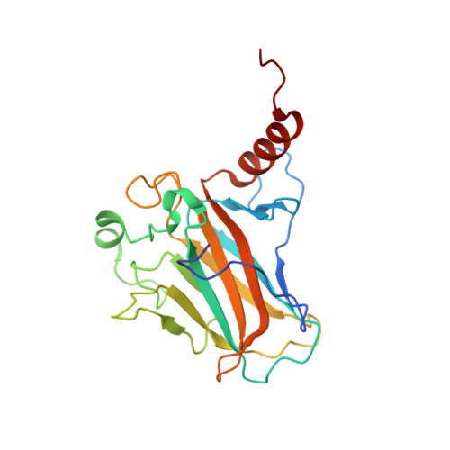
The 25-kDa core domain of the tumor suppressor p53 is inherently unstable and melts at just above body temperature, which makes it susceptible to oncogenic mutations that inactivate it by lowering its stability. We determined its structure in solution using state-of-the-art isotopic labeling techniques and NMR spectroscopy to complement its crystal structure. The structure was very similar to that in the crystal but far more mobile than expected. Importantly, we were able to analyze by NMR the structural environment of several buried polar groups, which indicated structural reasons for the instability. NMR spectroscopy, with its ability to detect protons, located buried hydroxyl and sulfhydryl groups that form suboptimal hydrogen-bond networks. We mutated one such buried pair, Tyr-236 and Thr-253 to Phe-236 and Ile-253 (as found in the paralogs p63 and p73), and stabilized p53 by 1.6 kcal/mol. We also detected differences in the conformation of a mobile loop that might reflect the existence of physiologically relevant alternative conformations. The effects of temperature on the dynamics of aromatic residues indicated that the protein also experiences several dynamic processes that might be related to the presence of alternative hydrogen-bond patterns in the protein interior. p53 appears to have evolved to be dynamic and unstable.
- Medical Research Council Centre for Protein Engineering, Medical Research Council Centre, Hills Road, Cambridge CB2 2QH, United Kingdom.
Organizational Affiliation: