2F5Z

Crystal Structure of Human Dihydrolipoamide Dehydrogenase (E3) Complexed to the E3-Binding Domain of Human E3-Binding Protein


Experimental Data Snapshot

  • Method: X-RAY DIFFRACTION
  • Resolution: 2.18 Å
  • R-Value Free: 0.242 
  • R-Value Work: 0.205 
  • R-Value Observed: 0.205 

wwPDB Validation   3D Report Full Report


Ligand Structure Quality Assessment 


This is version 1.4 of the entry. See complete history


Literature

Structural Insight into Interactions between Dihydrolipoamide Dehydrogenase (E3) and E3 Binding Protein of Human Pyruvate Dehydrogenase Complex.

Brautigam, C.A.Wynn, R.M.Chuang, J.L.Machius, M.Tomchick, D.R.Chuang, D.T.

(2006) Structure 14: 611-621

  • DOI: https://doi.org/10.1016/j.str.2006.01.001
  • Primary Citation of Related Structures:  
    2F5Z, 2F60

  • PubMed Abstract: 

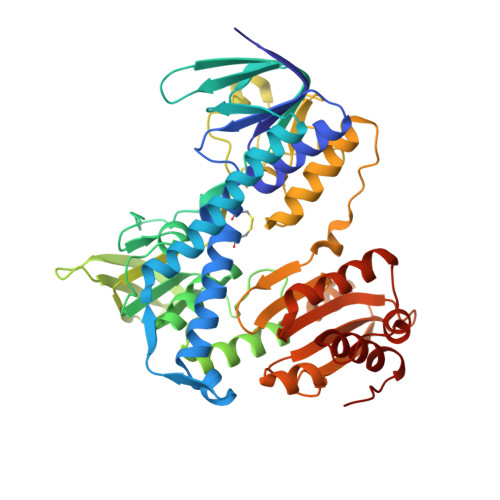
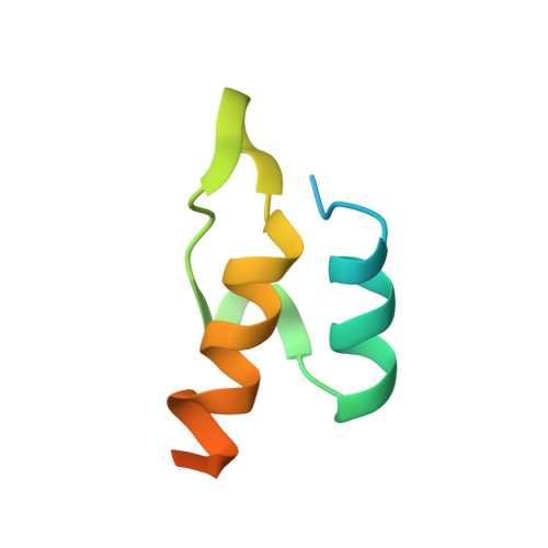
    The 9.5 MDa human pyruvate dehydrogenase complex (PDC) utilizes the specific dihydrolipoamide dehydrogenase (E3) binding protein (E3BP) to tether the essential E3 component to the 60-meric core of the complex. Here, we report crystal structures of the binding domain (E3BD) of human E3BP alone and in complex with human E3 at 1.6 angstroms and 2.2 angstroms, respectively. The latter structure shows that residues from E3BD contact E3 across its 2-fold axis, resulting in one E3BD binding site on the E3 homodimer. Negligible conformational changes occur in E3BD upon its high-affinity binding to E3. Modifications of E3BD residues at the center of the E3BD/E3 interface impede E3 binding far more severely than those of residues on the periphery, validating the "hot spot" paradigm for protein interactions. A cluster of disease-causing E3 mutations located near the center of the E3BD/E3 interface prevents the efficient recruitment of these E3 variants by E3BP into the PDC, leading to the dysfunction of the PDC catalytic machine.


  • Organizational Affiliation

    Department of Biochemistry, The University of Texas Southwestern Medical Center at Dallas, Dallas, Texas 75390, USA. chad.brautigam@utsouthwestern.edu


Macromolecules
Find similar proteins by:  (by identity cutoff)  |  3D Structure
Entity ID: 1
MoleculeChains Sequence LengthOrganismDetailsImage
Dihydrolipoyl dehydrogenase
A, B, C, D, E
A, B, C, D, E, F, G, H, I, J
474Homo sapiensMutation(s): 0 
Gene Names: DLDGCSLLADPHE3
EC: 1.8.1.4
UniProt & NIH Common Fund Data Resources
Find proteins for P09622 (Homo sapiens)
Explore P09622 
Go to UniProtKB:  P09622
PHAROS:  P09622
GTEx:  ENSG00000091140 
Entity Groups  
Sequence Clusters30% Identity50% Identity70% Identity90% Identity95% Identity100% Identity
UniProt GroupP09622
Sequence Annotations
Expand
  • Reference Sequence
Find similar proteins by:  (by identity cutoff)  |  3D Structure
Entity ID: 2
MoleculeChains Sequence LengthOrganismDetailsImage
Pyruvate dehydrogenase protein X component
K, L, M, N, O
64Homo sapiensMutation(s): 2 
Gene Names: PDHXPDX1
UniProt & NIH Common Fund Data Resources
Find proteins for O00330 (Homo sapiens)
Explore O00330 
Go to UniProtKB:  O00330
PHAROS:  O00330
GTEx:  ENSG00000110435 
Entity Groups  
Sequence Clusters30% Identity50% Identity70% Identity90% Identity95% Identity100% Identity
UniProt GroupO00330
Sequence Annotations
Expand
  • Reference Sequence
Small Molecules
Ligands 2 Unique
IDChains Name / Formula / InChI Key2D Diagram3D Interactions
FAD
Query on FAD

Download Ideal Coordinates CCD File 
CB [auth H]
DA [auth C]
GB [auth I]
IA [auth D]
LB [auth J]
CB [auth H],
DA [auth C],
GB [auth I],
IA [auth D],
LB [auth J],
NA [auth E],
SA [auth F],
T [auth A],
XA [auth G],
Y [auth B]
FLAVIN-ADENINE DINUCLEOTIDE
C27 H33 N9 O15 P2
VWWQXMAJTJZDQX-UYBVJOGSSA-N
SO4
Query on SO4

Download Ideal Coordinates CCD File 
AA [auth C]
AB [auth H]
BA [auth C]
BB [auth H]
CA [auth C]
AA [auth C],
AB [auth H],
BA [auth C],
BB [auth H],
CA [auth C],
DB [auth I],
EA [auth D],
EB [auth I],
FA [auth D],
FB [auth I],
GA [auth D],
HA [auth D],
HB [auth J],
IB [auth J],
JA [auth E],
JB [auth J],
KA [auth E],
KB [auth J],
LA [auth E],
MA [auth E],
OA [auth F],
P [auth A],
PA [auth F],
Q [auth A],
QA [auth F],
R [auth A],
RA [auth F],
S [auth A],
TA [auth G],
U [auth B],
UA [auth G],
V [auth B],
VA [auth G],
W [auth B],
WA [auth G],
X [auth B],
YA [auth H],
Z [auth C],
ZA [auth H]
SULFATE ION
O4 S
QAOWNCQODCNURD-UHFFFAOYSA-L
Experimental Data & Validation

Experimental Data

  • Method: X-RAY DIFFRACTION
  • Resolution: 2.18 Å
  • R-Value Free: 0.242 
  • R-Value Work: 0.205 
  • R-Value Observed: 0.205 
  • Space Group: P 21 21 21
Unit Cell:
Length ( Å )Angle ( ˚ )
a = 171.153α = 90
b = 187.728β = 90
c = 224.392γ = 90
Software Package:
Software NamePurpose
HKL-2000data collection
SCALEPACKdata scaling
CNSrefinement
HKL-2000data reduction
CNSphasing

Structure Validation

View Full Validation Report


Ligand Structure Quality Assessment 


Entry History 

Deposition Data

Revision History  (Full details and data files)

  • Version 1.0: 2006-01-17
    Type: Initial release
  • Version 1.1: 2008-05-01
    Changes: Version format compliance
  • Version 1.2: 2011-07-13
    Changes: Version format compliance
  • Version 1.3: 2021-10-20
    Changes: Database references, Derived calculations
  • Version 1.4: 2023-08-23
    Changes: Data collection, Refinement description