2DKE | pdb_00002dke

Crystal structure of substrate-free form of PcyA


Experimental Data Snapshot

  • Method: X-RAY DIFFRACTION
  • Resolution: 2.50 Å
  • R-Value Free: 
    0.299 (Depositor), 0.230 (DCC) 
  • R-Value Work: 
    0.243 (Depositor), 0.244 (DCC) 
  • R-Value Observed: 
    0.248 (Depositor) 

Starting Model: experimental
View more details

wwPDB Validation 3D Report Full Report

Validation slider image for 2DKE

This is version 1.3 of the entry. See complete history

Literature

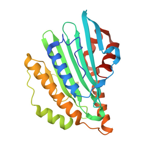
Induced-fitting and electrostatic potential change of PcyA upon substrate binding demonstrated by the crystal structure of the substrate-free form

Hagiwara, Y.Sugishima, M.Takahashi, Y.Fukuyama, K.

(2006) FEBS Lett 580: 3823-3828

  • DOI: https://doi.org/10.1016/j.febslet.2006.05.075
  • Primary Citation Related Structures: 
    2DKE

  • PubMed Abstract: 

    Phycocyanobilin:ferredoxin oxidoreductase (PcyA) catalyzes the sequential reduction of the vinyl group of the D-ring and the A-ring of biliverdin IXalpha (BV) using ferredoxin to produce phycocyanobilin, a pigment used for light-harvesting and light-sensing in red algae and cyanobacteria. We have determined the crystal structure of the substrate-free form of PcyA from Synechocystis sp. PCC 6803 at 2.5 A resolution. Structural comparison of the substrate-free form and the PcyA-BV complex shows major changes around the entrance of the BV binding pocket; upon BV binding, two alpha-helices and nearby side-chains move to produce tight BV binding. Unexpectedly, these movements localize the positive charges around the BV binding site, which may contribute to the proper binding of ferredoxin to PcyA. In the substrate-free form, the side-chain of Asp105 was located at a site that would be underneath the BV A-ring in the PcyA-BV complex and hydrogen-bonded with His88. We propose that BV is protonated by a mechanism involving conformational changes of these two residues before reduction.


  • Organizational Affiliation
    • Department of Biological Sciences, Graduate School of Science, Osaka University, 1-1 Machikaneyama-cho, Toyonaka, Osaka 560-0043, Japan.

Macromolecule Content 

  • Total Structure Weight: 28.19 kDa 
  • Atom Count: 1,970 
  • Modeled Residue Count: 241 
  • Deposited Residue Count: 248 
  • Unique protein chains: 1

Macromolecules

Find similar proteins by:|  3D Structure
Entity ID: 1
MoleculeChains  Sequence LengthOrganismDetailsImage
Phycocyanobilin:ferredoxin oxidoreductase248Synechocystis sp. PCC 6803Mutation(s): 0 
EC: 1.3.7.5
UniProt
Find proteins for Q55891 (Synechocystis sp. (strain ATCC 27184 / PCC 6803 / Kazusa))
Explore Q55891 
Go to UniProtKB:  Q55891
Entity Groups
Sequence Clusters30% Identity50% Identity70% Identity90% Identity95% Identity100% Identity
UniProt GroupQ55891
Sequence Annotations
Expand
Reference Sequence

Small Molecules

Ligands 1 Unique
IDChains Name / Formula / InChI Key2D Diagram3D Interactions
CL

Query on CL


Download:Ideal Coordinates CCD File
B [auth A]CHLORIDE ION
Cl
VEXZGXHMUGYJMC-UHFFFAOYSA-M

Experimental Data & Validation

Experimental Data

  • Method: X-RAY DIFFRACTION
  • Resolution: 2.50 Å
  • R-Value Free:  0.299 (Depositor), 0.230 (DCC) 
  • R-Value Work:  0.243 (Depositor), 0.244 (DCC) 
  • R-Value Observed: 0.248 (Depositor) 
Space Group: P 43 21 2
Unit Cell:
Length ( Å )Angle ( ˚ )
a = 75.98α = 90
b = 75.98β = 90
c = 84.97γ = 90
Software Package:
Software NamePurpose
HKL-2000data collection
SCALEPACKdata scaling
MOLREPphasing
CNSrefinement
HKL-2000data reduction

Structure Validation

View Full Validation Report


Entry History 

Deposition Data

Revision History  (Full details and data files)

  • Version 1.0: 2006-07-25
    Type: Initial release
  • Version 1.1: 2008-04-30
    Changes: Version format compliance
  • Version 1.2: 2011-07-13
    Changes: Source and taxonomy, Version format compliance
  • Version 1.3: 2023-10-25
    Changes: Data collection, Database references, Derived calculations, Refinement description