2D4M

Crystal Structure of apo M-PMV dUTPase


Experimental Data Snapshot

  • Method: X-RAY DIFFRACTION
  • Resolution: 1.85 Å
  • R-Value Free: 0.192 
  • R-Value Work: 0.160 
  • R-Value Observed: 0.162 

wwPDB Validation   3D Report Full Report


This is version 1.7 of the entry. See complete history


Literature

Flexible segments modulate co-folding of dUTPase and nucleocapsid proteins.

Nemeth-Pongracz, V.Barabas, O.Fuxreiter, M.Simon, I.Pichova, I.Rumlova, M.Zabranska, H.Svergun, D.Petoukhov, M.Harmat, V.Klement, E.Hunyadi-Gulyas, E.Medzihradszky, K.F.Konya, E.Vertessy, B.G.

(2007) Nucleic Acids Res 35: 495-505

  • DOI: https://doi.org/10.1093/nar/gkl1074
  • Primary Citation of Related Structures:  
    2D4L, 2D4M, 2D4N

  • PubMed Abstract: 

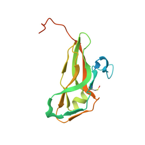
    The homotrimeric fusion protein nucleocapsid (NC)-dUTPase combines domains that participate in RNA/DNA folding, reverse transcription, and DNA repair in Mason-Pfizer monkey betaretrovirus infected cells. The structural organization of the fusion protein remained obscured by the N- and C-terminal flexible segments of dUTPase and the linker region connecting the two domains that are invisible in electron density maps. Small-angle X-ray scattering reveals that upon oligonucleotide binding the NC domains adopt the trimeric symmetry of dUTPase. High-resolution X-ray structures together with molecular modeling indicate that fusion with NC domains dramatically alters the conformation of the flexible C-terminus by perturbing the orientation of a critical beta-strand. Consequently, the C-terminal segment is capable of double backing upon the active site of its own monomer and stabilized by non-covalent interactions formed with the N-terminal segment. This co-folding of the dUTPase terminal segments, not observable in other homologous enzymes, is due to the presence of the fused NC domain. Structural and genomic advantages of fusing the NC domain to a shortened dUTPase in betaretroviruses and the possible physiological consequences are envisaged.


  • Organizational Affiliation

    Institute of Enzymology, BRC, Hungarian Academy of Sciences, Budapest, Karolina út 29, H-1113, Hungary.


Macromolecules
Find similar proteins by:  (by identity cutoff)  |  3D Structure
Entity ID: 1
MoleculeChains Sequence LengthOrganismDetailsImage
DU152Mason-Pfizer monkey virusMutation(s): 1 
Gene Names: gag-pro
EC: 3.6.1.23
UniProt
Find proteins for P07570 (Mason-Pfizer monkey virus)
Explore P07570 
Go to UniProtKB:  P07570
Entity Groups  
Sequence Clusters30% Identity50% Identity70% Identity90% Identity95% Identity100% Identity
UniProt GroupP07570
Sequence Annotations
Expand
  • Reference Sequence
Small Molecules
Modified Residues  1 Unique
IDChains TypeFormula2D DiagramParent
CSO
Query on CSO
A
L-PEPTIDE LINKINGC3 H7 N O3 SCYS
Experimental Data & Validation

Experimental Data

  • Method: X-RAY DIFFRACTION
  • Resolution: 1.85 Å
  • R-Value Free: 0.192 
  • R-Value Work: 0.160 
  • R-Value Observed: 0.162 
  • Space Group: P 63
Unit Cell:
Length ( Å )Angle ( ˚ )
a = 61.191α = 90
b = 61.191β = 90
c = 63.31γ = 120
Software Package:
Software NamePurpose
REFMACrefinement
MAR345data collection
XDSdata scaling

Structure Validation

View Full Validation Report


Entry History 

Deposition Data

Revision History  (Full details and data files)

  • Version 1.0: 2006-11-21
    Type: Initial release
  • Version 1.1: 2008-04-30
    Changes: Version format compliance
  • Version 1.2: 2011-07-13
    Changes: Derived calculations, Version format compliance
  • Version 1.3: 2011-07-20
    Changes: Database references
  • Version 1.4: 2017-10-11
    Changes: Refinement description
  • Version 1.5: 2021-11-10
    Changes: Data collection, Database references, Derived calculations
  • Version 1.6: 2023-10-25
    Changes: Data collection, Refinement description
  • Version 1.7: 2023-11-15
    Changes: Data collection