2D2Q

Crystal structure of the dimerized radixin FERM domain


Experimental Data Snapshot

  • Method: X-RAY DIFFRACTION
  • Resolution: 2.80 Å
  • R-Value Free: 0.269 
  • R-Value Work: 0.222 

wwPDB Validation   3D Report Full Report


This is version 1.3 of the entry. See complete history


Literature

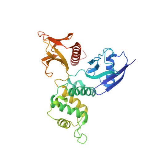
Structure of dimerized radixin FERM domain suggests a novel masking motif in C-terminal residues 295-304

Kitano, K.Yusa, F.Hakoshima, T.

(2006) Acta Crystallogr Sect F Struct Biol Cryst Commun 62: 340-345

  • DOI: https://doi.org/10.1107/S1744309106010062
  • Primary Citation of Related Structures:  
    2D2Q

  • PubMed Abstract: 

    ERM (ezrin/radixin/moesin) proteins bind to the cytoplasmic tail of adhesion molecules in the formation of the membrane-associated cytoskeleton. The binding site is located in the FERM (4.1 and ERM) domain, a domain that is masked in the inactive form. A conventional masking motif, strand 1 (residues 494-500 in radixin), has previously been identified in the C-terminal tail domain. Here, the crystal structure of dimerized radixin FERM domains (residues 1-310) is presented in which the binding site of one molecule is occupied by the C-terminal residues (residues 295-304, strand 2) of the other molecule. The residues contain a conserved motif that is compatible with that identified in the adhesion molecules. The residues might serve as a second masking region in the inactive form of ERM proteins.


  • Organizational Affiliation

    Structural Biology Laboratory, Nara Institute of Science and Technology, 8916-5 Takayama, Ikoma, Nara 630-0192, Japan.


Macromolecules
Find similar proteins by:  (by identity cutoff)  |  3D Structure
Entity ID: 1
MoleculeChains Sequence LengthOrganismDetailsImage
Radixin
A, B
310Mus musculusMutation(s): 0 
UniProt & NIH Common Fund Data Resources
Find proteins for P26043 (Mus musculus)
Explore P26043 
Go to UniProtKB:  P26043
IMPC:  MGI:97887
Entity Groups  
Sequence Clusters30% Identity50% Identity70% Identity90% Identity95% Identity100% Identity
UniProt GroupP26043
Sequence Annotations
Expand
  • Reference Sequence
Experimental Data & Validation

Experimental Data

  • Method: X-RAY DIFFRACTION
  • Resolution: 2.80 Å
  • R-Value Free: 0.269 
  • R-Value Work: 0.222 
  • Space Group: P 1 21 1
Unit Cell:
Length ( Å )Angle ( ˚ )
a = 57.218α = 90
b = 70.218β = 98.97
c = 110.81γ = 90
Software Package:
Software NamePurpose
HKL-2000data collection
SCALEPACKdata scaling
CNSrefinement
HKL-2000data reduction
CNSphasing

Structure Validation

View Full Validation Report


Entry History 

Deposition Data

Revision History  (Full details and data files)

  • Version 1.0: 2006-04-18
    Type: Initial release
  • Version 1.1: 2008-04-30
    Changes: Version format compliance
  • Version 1.2: 2011-07-13
    Changes: Version format compliance
  • Version 1.3: 2024-03-13
    Changes: Data collection, Database references