Crystallographic snapshots of an entire reaction cycle for a retaining xylanase from Streptomyces olivaceoviridis E-86
Suzuki, R., Fujimoto, Z., Ito, S., Kawahara, S., Kaneko, S., Taira, K., Hasegawa, T., Kuno, A.(2009) J Biochem 146: 61-70
- PubMed: 19279191 Search on PubMed
- DOI: https://doi.org/10.1093/jb/mvp047
- Primary Citation Related Structures:
2D1Z, 2D20, 2D22, 2D23, 2D24 - PubMed Abstract:
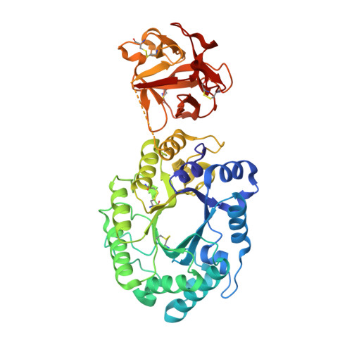
Retaining glycosyl hydrolases, which catalyse both glycosylation and deglycosylation in a concerted manner, are the most abundant hydrolases. To date, their visualization has tended to be focused on glycosylation because glycosylation reactions can be visualized by inactivating deglycosylation step and/or using substrate analogues to isolate covalent intermediates. Furthermore, during structural analyses of glycosyl hydrolases with hydrolytic reaction products by the conventional soaking method, mutarotation of an anomeric carbon in the reaction products promptly and certainly occurs. This undesirable structural alteration hinders visualization of the second step in the reaction. Here, we investigated X-ray crystallographic visualization as a possible method for visualizing the conformational itinerary of a retaining xylanase from Streptomyces olivaceoviridis E-86. To clearly define the stereochemistry at the anomeric carbon during the deglycosylation step, extraneous nucleophiles, such as azide, were adopted to substitute for the missing base catalyst in an appropriate mutant. The X-ray crystallographic visualization provided snapshots of the components of the entire reaction, including the E*S complex, the covalent intermediate, breakdown of the intermediate and the enzyme-product (E*P)complex.
- Department of Material and Biological Chemistry, Yamagata University, Japan.
Organizational Affiliation: