2CQT

Crystal Structure of Cellvibrio gilvus Cellobiose Phosphorylase Crystallized from Sodium/Potassium Phosphate


Experimental Data Snapshot

  • Method: X-RAY DIFFRACTION
  • Resolution: 2.10 Å
  • R-Value Free: 0.220 
  • R-Value Work: 0.180 
  • R-Value Observed: 0.180 

wwPDB Validation   3D Report Full Report


Ligand Structure Quality Assessment 


This is version 1.4 of the entry. See complete history


Literature

Structural dissection of the reaction mechanism of cellobiose phosphorylase.

Hidaka, M.Kitaoka, M.Hayashi, K.Wakagi, T.Shoun, H.Fushinobu, S.

(2006) Biochem J 398: 37-43

  • DOI: https://doi.org/10.1042/BJ20060274
  • Primary Citation of Related Structures:  
    2CQS, 2CQT

  • PubMed Abstract: 

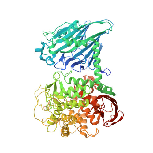
    Cellobiose phosphorylase, a member of the glycoside hydrolase family 94, catalyses the reversible phosphorolysis of cellobiose into alpha-D-glucose 1-phosphate and D-glucose with inversion of the anomeric configuration. The substrate specificity and reaction mechanism of cellobiose phosphorylase from Cellvibrio gilvus have been investigated in detail. We have determined the crystal structure of the glucose-sulphate and glucose-phosphate complexes of this enzyme at a maximal resolution of 2.0 A (1 A=0.1 nm). The phosphate ion is strongly held through several hydrogen bonds, and the configuration appears to be suitable for direct nucleophilic attack to an anomeric centre. Structural features around the sugar-donor and sugar-acceptor sites were consistent with the results of extensive kinetic studies. When we compared this structure with that of homologous chitobiose phosphorylase, we identified key residues for substrate discrimination between glucose and N-acetylglucosamine in both the sugar-donor and sugar-acceptor sites. We found that the active site pocket of cellobiose phosphorylase was covered by an additional loop, indicating that some conformational change is required upon substrate binding. Information on the three-dimensional structure of cellobiose phosphorylase will facilitate engineering of this enzyme, the application of which to practical oligosaccharide synthesis has already been established.


  • Organizational Affiliation

    Department of Biotechnology, University of Tokyo, 1-1-1 Yayoi, Bunkyo-ku, Tokyo 113-8657, Japan.


Macromolecules
Find similar proteins by:  (by identity cutoff)  |  3D Structure
Entity ID: 1
MoleculeChains Sequence LengthOrganismDetailsImage
Cellobiose Phosphorylase
A, B
842Cellulomonas gilvusMutation(s): 0 
EC: 2.4.1.20
UniProt
Find proteins for O66264 (Cellulomonas gilvus)
Explore O66264 
Go to UniProtKB:  O66264
Entity Groups  
Sequence Clusters30% Identity50% Identity70% Identity90% Identity95% Identity100% Identity
UniProt GroupO66264
Sequence Annotations
Expand
  • Reference Sequence
Experimental Data & Validation

Experimental Data

  • Method: X-RAY DIFFRACTION
  • Resolution: 2.10 Å
  • R-Value Free: 0.220 
  • R-Value Work: 0.180 
  • R-Value Observed: 0.180 
  • Space Group: P 1 21 1
Unit Cell:
Length ( Å )Angle ( ˚ )
a = 85.275α = 90
b = 98.8β = 102.53
c = 105.102γ = 90
Software Package:
Software NamePurpose
CNSrefinement
HKL-2000data reduction
SCALEPACKdata scaling
MOLREPphasing

Structure Validation

View Full Validation Report


Ligand Structure Quality Assessment 


Entry History 

Deposition Data

Revision History  (Full details and data files)

  • Version 1.0: 2006-05-16
    Type: Initial release
  • Version 1.1: 2008-04-30
    Changes: Version format compliance
  • Version 1.2: 2011-07-13
    Changes: Non-polymer description, Version format compliance
  • Version 1.3: 2020-07-29
    Type: Remediation
    Reason: Carbohydrate remediation
    Changes: Data collection, Database references, Derived calculations, Structure summary
  • Version 1.4: 2023-10-25
    Changes: Data collection, Database references, Refinement description, Structure summary