2CPO | pdb_00002cpo

CHLOROPEROXIDASE


Experimental Data Snapshot

  • Method: X-RAY DIFFRACTION
  • Resolution: 2.10 Å
  • R-Value Free: 
    0.228 (Depositor) 
  • R-Value Work: 
    0.186 (Depositor), 0.179 (DCC) 
  • R-Value Observed: 
    0.186 (Depositor) 

wwPDB Validation 3D Report Full Report

Validation slider image for 2CPO

Ligand Structure Quality Assessment 


This is version 3.1 of the entry. See complete history

Literature

The crystal structure of chloroperoxidase: a heme peroxidase--cytochrome P450 functional hybrid.

Sundaramoorthy, M.Terner, J.Poulos, T.L.

(1995) Structure 3: 1367-1377

  • DOI: https://doi.org/10.1016/s0969-2126(01)00274-x
  • Primary Citation Related Structures: 
    1CPO, 2CPO

  • PubMed Abstract: 

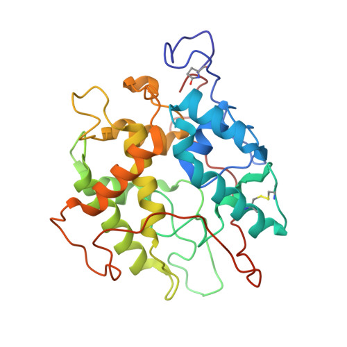
    Chloroperoxidase (CPO) is a versatile heme-containing enzyme that exhibits peroxidase, catalase and cytochrome P450-like activities in addition to catalyzing halogenation reactions. The structure determination of CPO was undertaken to help elucidate those structural features that enable the enzyme to exhibit these multiple activities. Despite functional similarities with other heme enzymes, CPO folds into a novel tertiary structure dominated by eight helical segments. The catalytic base, required to cleave the peroxide O-O bond, is glutamic acid rather than histidine as in other peroxidases. CPO contains a hydrophobic patch above the heme that could be the binding site for substrates that undergo P450-like reactions. The crystal structure also shows extensive glycosylation with both N- and O-linked glycosyl chains. The proximal side of the heme in CPO resembles cytochrome P450 because a cysteine residue serves as an axial heme ligand, whereas the distal side of the heme is 'peroxidase-like' in that polar residues form the peroxide-binding site. Access to the heme pocket is restricted to the distal face such that small organic substrates can interact with the iron-linked oxygen atom which accounts for the P450-like reactions catalyzed by chloroperoxidase.


  • Organizational Affiliation
    • Department of Molecular Biology and Biochemistry, University of California, Irvine 92717, USA.

Macromolecule Content 

  • Total Structure Weight: 35.93 kDa 
  • Atom Count: 2,690 
  • Modeled Residue Count: 299 
  • Deposited Residue Count: 299 
  • Unique protein chains: 1

Macromolecules

Find similar proteins by:|  3D Structure
Entity ID: 1
MoleculeChains  Sequence LengthOrganismDetailsImage
CHLOROPEROXIDASE299Leptoxyphium fumagoMutation(s): 0 
EC: 1.11.1.10
UniProt
Find proteins for P04963 (Leptoxyphium fumago)
Explore P04963 
Go to UniProtKB:  P04963
Entity Groups
Sequence Clusters30% Identity50% Identity70% Identity90% Identity95% Identity100% Identity
UniProt GroupP04963
Glycosylation
Glycosylation Sites: 11
Sequence Annotations
Expand
Reference Sequence

Oligosaccharides

Help  
Entity ID: 2
MoleculeChains Length2D Diagram GlycosylationD Interactions
2-acetamido-2-deoxy-beta-D-glucopyranose-(1-4)-2-acetamido-2-deoxy-beta-D-glucopyranose
B, C
2N-Glycosylation
Glycosylation Resources
GlyTouCan: G42666HT
GlyCosmos: G42666HT
GlyGen: G42666HT

Small Molecules

Ligands 4 Unique
IDChains Name / Formula / InChI Key2D Diagram3D Interactions
HEM

Query on HEM


Download:Ideal Coordinates CCD File
N [auth A]PROTOPORPHYRIN IX CONTAINING FE
C34 H32 Fe N4 O4
KABFMIBPWCXCRK-RGGAHWMASA-L
NAG

Query on NAG


Download:Ideal Coordinates CCD File
D [auth A]2-acetamido-2-deoxy-beta-D-glucopyranose
C8 H15 N O6
OVRNDRQMDRJTHS-FMDGEEDCSA-N
MAN

Query on MAN


Download:Ideal Coordinates CCD File
E [auth A]
F [auth A]
G [auth A]
H [auth A]
I [auth A]
E [auth A],
F [auth A],
G [auth A],
H [auth A],
I [auth A],
J [auth A],
K [auth A],
L [auth A]
alpha-D-mannopyranose
C6 H12 O6
WQZGKKKJIJFFOK-PQMKYFCFSA-N
MN

Query on MN


Download:Ideal Coordinates CCD File
M [auth A]MANGANESE (II) ION
Mn
WAEMQWOKJMHJLA-UHFFFAOYSA-N
Modified Residues  1 Unique
IDChains TypeFormula2D DiagramParent
PCA
Query on PCA
A
L-PEPTIDE LINKINGC5 H7 N O3GLN

Experimental Data & Validation

Experimental Data

  • Method: X-RAY DIFFRACTION
  • Resolution: 2.10 Å
  • R-Value Free:  0.228 (Depositor) 
  • R-Value Work:  0.186 (Depositor), 0.179 (DCC) 
  • R-Value Observed: 0.186 (Depositor) 
Space Group: C 2 2 21
Unit Cell:
Length ( Å )Angle ( ˚ )
a = 58.62α = 90
b = 152.17β = 90
c = 102.1γ = 90
Software Package:
Software NamePurpose
XENGENdata collection
X-PLORmodel building
X-PLORrefinement
XENGENdata reduction
X-PLORphasing

Structure Validation

View Full Validation Report


Ligand Structure Quality Assessment 


Entry History 

Deposition Data

Revision History  (Full details and data files)

  • Version 1.0: 1997-02-12
    Type: Initial release
  • Version 1.1: 2008-03-24
    Changes: Version format compliance
  • Version 1.2: 2011-07-13
    Changes: Non-polymer description, Version format compliance
  • Version 2.0: 2019-12-25
    Changes: Derived calculations, Other, Polymer sequence
  • Version 3.0: 2020-07-29
    Type: Remediation
    Reason: Carbohydrate remediation
    Changes: Advisory, Atomic model, Data collection, Derived calculations, Structure summary
  • Version 3.1: 2024-10-30
    Changes: Data collection, Database references, Structure summary