Spectroscopic Characterization of Successive Phosphorylation of the Tissue Factor Cytoplasmic Region.
Sen, M., Herzik, M., Craft, J.W., Creath, A.L., Agrawal, S., Ruf, W., Legge, G.B.(2009) Open Spectrosc J 3: 58
- PubMed: 20076769 Search on PubMedSearch on PubMed Central
- DOI: https://doi.org/10.2174/1874383800903010058
- Primary Citation Related Structures:
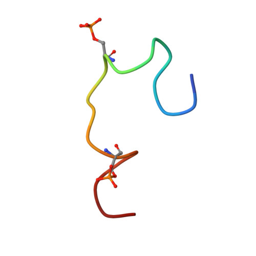
2CEF, 2CEH, 2CEZ, 2CFJ - PubMed Abstract:
Tissue Factor (TF) is well known for its role during the activation of the coagulation pathway, but it is also critical for tumor biology and inflammation through protease activated receptor (PAR) 2 signaling. This signaling function is modulated by the successive phosphorylation of residues Ser253 and Ser258 within the TF cytoplasmic region (TFCR). This paper reports how we used NMR and spectroscopic methods to investigate the structural propensities of the unphosphorylated and phosphorylated forms of the TFCR. When unphosphorylated, the TFCR forms a local hydrophobic collapse around Trp254 and an electropositive patch from the membrane proximal basic block (Arg246-Lys247) to the conserved PKCalpha consensus residue Lys255. Phosphorylation of Ser253 alters the charge characteristics of this membrane proximal region, thereby strengthening the interaction between residue Ala248 and the Trp254 aromatic group. Phosphorylation of the Ser258-Pro259 motif destabilizes a turn at the C-terminus to form an extended polyproline helical motif. Our data suggests that by changing both its charge and local structural propensity, covalent modifications of the TFCR can potentially regulate its association with the cellular membrane and its signaling partners.
- University of Houston, Department of Biology and Biochemistry, Houston, TX 77204-5001, USA.
Organizational Affiliation: