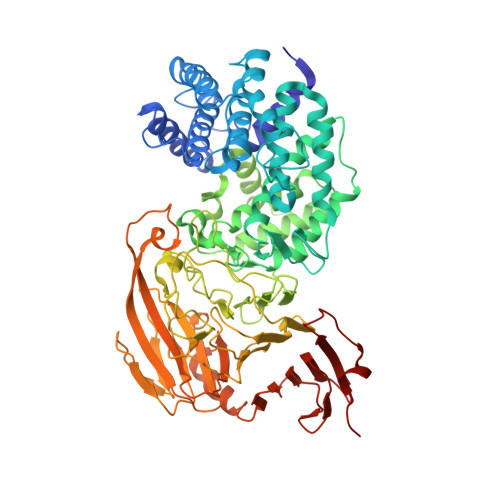
Alternate Structural Conformations of Streptococcus Pneumoniae Hyaluronan Lyase: Insights Into Enzyme Flexibility and Underlying Molecular Mechanism of Action.
Rigden, D.J., Littlejohn, J.E., Joshi, H.V., De Groot, B.L., Jedrzejas, M.J.(2006) J Mol Biology 358: 1165
- PubMed: 16569416 Search on PubMed
- DOI: https://doi.org/10.1016/j.jmb.2006.02.066
- Primary Citation Related Structures:
2BRV, 2BRW - PubMed Abstract:
Streptococcus pneumoniae hyaluronan lyase is a surface enzyme of this Gram-positive bacterium. The enzyme degrades several biologically important, information-rich linear polymeric glycans: hyaluronan, unsulfated chondroitin, and some chondroitin sulfates. This degradation facilitates spreading of bacteria throughout the host tissues and presumably provides energy and a carbon source for pneumococcal cells. Its beta-elimination catalytic mechanism is an acid/base process termed proton acceptance and donation leading to cleavage of beta-1,4 linkages of the substrates. The degradation of hyaluronan occurs in two stages, initial endolytic cuts are followed by processive exolytic cleavage of one disaccharide at a time. In contrast, the degradation of chondroitins is purely endolytic. Structural studies together with flexibility analyses of two streptococcal enzymes, from S.pneumoniae and Streptococcus agalactiae, allowed for insights into this enzyme's molecular mechanism. Here, two new X-ray crystal structures of the pneumococcal enzyme in novel conformations are reported. These new conformations, complemented by molecular dynamics simulation results, directly confirm the predicted domain motions presumed to facilitate the processive degradative process. One of these new structures resembles the S.agalactiae enzyme conformation, and provides evidence of a uniform mechanistic/dynamic behavior of this protein across different bacteria.
- Children's Hospital Oakland Research Institute, Oakland, CA 94609, USA.
Organizational Affiliation: