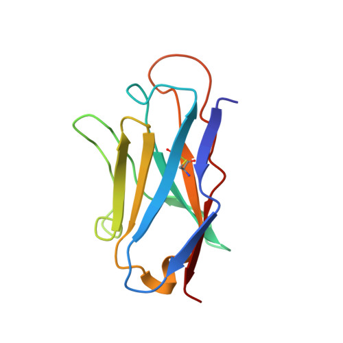
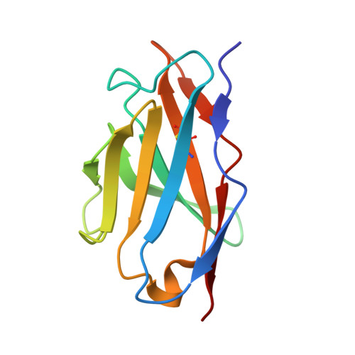
2BJM

SPE7:Anthrone Complex


Experimental Data Snapshot

  • Method: X-RAY DIFFRACTION
  • Resolution: 2.15 Å
  • R-Value Free: 0.286 
  • R-Value Work: 0.268 
  • R-Value Observed: 0.269 

wwPDB Validation   3D Report Full Report


This is version 1.1 of the entry. See complete history


Literature

Structure and Kinetics of a Transient Antibody Binding Intermediate Reveal a Kinetic Discrimination Mechanism in Antigen Recognition

James, L.C.Tawfik, D.S.

(2005) Proc Natl Acad Sci U S A 102: 12730

  • DOI: https://doi.org/10.1073/pnas.0500909102
  • Primary Citation of Related Structures:  
    2BJM

  • PubMed Abstract: 

    Induced fit is a predominant phenomenon in protein-ligand interactions, yet it is invariably attributed without establishing the existence, let alone the structure, of the initial, low-affinity encounter complex. We determined the crystal structure of the encounter complex on the pathway of ligand binding by IgE antibody SPE7. We show that this complex is formed by a wide range of ligands that initially bind with identical affinity. Nonspecific ligands rapidly dissociate, whereupon the antibody isomerizes to a nonbinding isomer. Specific ligand complexes, however, slowly isomerize to give a high-affinity complex. This isomerization involves backbone and side-chain rearrangements of up to 14 A and the formation of specific hydrogen bonds. The postbinding conformational switch, combined with the prebinding isomerization to an energetically favorable nonbinding isomer, results in a "kinetic discrimination" mechanism that mediates selective binding, by a factor of >10(3), between highly related ligands that initially bind with the same affinity. This model may apply to proteins that bind multiple ligands in a specific manner or other proteins that, although capable of binding many ligands, are activated by only a few.


  • Organizational Affiliation

    Laboratory of Molecular Biology, Medical Research Council, Cambridge CB2 2HQ, United Kingdom. lcj@mrc-lmb.cam.ac.uk


Macromolecules
Find similar proteins by:  (by identity cutoff)  |  3D Structure
Entity ID: 1
MoleculeChains Sequence LengthOrganismDetailsImage
IGE SPE7 HEAVY CHAINA [auth H]120Mus musculusMutation(s): 0 
Entity Groups  
Sequence Clusters30% Identity50% Identity70% Identity90% Identity95% Identity100% Identity
Sequence Annotations
Expand
  • Reference Sequence
Find similar proteins by:  (by identity cutoff)  |  3D Structure
Entity ID: 2
MoleculeChains Sequence LengthOrganismDetailsImage
IGE SPE7 LIGHT CHAINB [auth L]110Mus musculusMutation(s): 0 
UniProt
Find proteins for P01724 (Mus musculus)
Explore P01724 
Go to UniProtKB:  P01724
Entity Groups  
Sequence Clusters30% Identity50% Identity70% Identity90% Identity95% Identity100% Identity
UniProt GroupP01724
Sequence Annotations
Expand
  • Reference Sequence
Small Molecules
Ligands 1 Unique
IDChains Name / Formula / InChI Key2D Diagram3D Interactions
ANF
Query on ANF

Download Ideal Coordinates CCD File 
C [auth H]ANTHRONE
C14 H10 O
AUKRYONWZHRJRE-UHFFFAOYSA-N
Binding Affinity Annotations 
IDSourceBinding Affinity
ANF PDBBind:  2BJM Kd: 2.00e+5 (nM) from 1 assay(s)
Binding MOAD:  2BJM Ka: 1.00e+5 (M^-1) from 1 assay(s)
Experimental Data & Validation

Experimental Data

  • Method: X-RAY DIFFRACTION
  • Resolution: 2.15 Å
  • R-Value Free: 0.286 
  • R-Value Work: 0.268 
  • R-Value Observed: 0.269 
  • Space Group: I 4
Unit Cell:
Length ( Å )Angle ( ˚ )
a = 79.672α = 90
b = 79.672β = 90
c = 67.958γ = 90
Software Package:
Software NamePurpose
REFMACrefinement

Structure Validation

View Full Validation Report


Entry History 

Deposition Data

Revision History  (Full details and data files)

  • Version 1.0: 2005-08-18
    Type: Initial release
  • Version 1.1: 2013-10-30
    Changes: Derived calculations, Other, Source and taxonomy, Structure summary, Version format compliance