2BJM | pdb_00002bjm

SPE7:Anthrone Complex


Experimental Data Snapshot

  • Method: X-RAY DIFFRACTION
  • Resolution: 2.15 Å
  • R-Value Free: 
    0.286 (Depositor), 0.341 (DCC) 
  • R-Value Work: 
    0.268 (Depositor), 0.345 (DCC) 
  • R-Value Observed: 
    0.269 (Depositor) 

wwPDB Validation 3D Report Full Report

Validation slider image for 2BJM

Ligand Structure Quality Assessment 


This is version 1.2 of the entry. See complete history

Literature

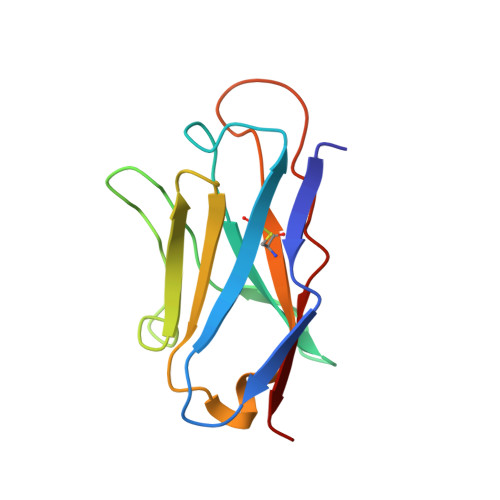
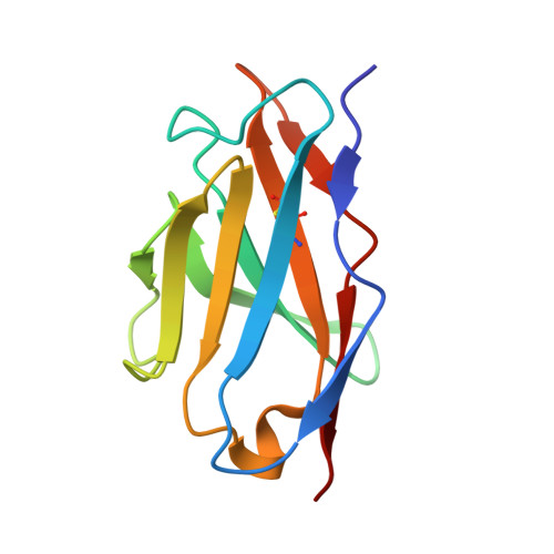
Structure and Kinetics of a Transient Antibody Binding Intermediate Reveal a Kinetic Discrimination Mechanism in Antigen Recognition

James, L.C.Tawfik, D.S.

(2005) Proc Natl Acad Sci U S A 102: 12730

  • DOI: https://doi.org/10.1073/pnas.0500909102
  • Primary Citation Related Structures: 
    2BJM

  • PubMed Abstract: 

    Induced fit is a predominant phenomenon in protein-ligand interactions, yet it is invariably attributed without establishing the existence, let alone the structure, of the initial, low-affinity encounter complex. We determined the crystal structure of the encounter complex on the pathway of ligand binding by IgE antibody SPE7. We show that this complex is formed by a wide range of ligands that initially bind with identical affinity. Nonspecific ligands rapidly dissociate, whereupon the antibody isomerizes to a nonbinding isomer. Specific ligand complexes, however, slowly isomerize to give a high-affinity complex. This isomerization involves backbone and side-chain rearrangements of up to 14 A and the formation of specific hydrogen bonds. The postbinding conformational switch, combined with the prebinding isomerization to an energetically favorable nonbinding isomer, results in a "kinetic discrimination" mechanism that mediates selective binding, by a factor of >10(3), between highly related ligands that initially bind with the same affinity. This model may apply to proteins that bind multiple ligands in a specific manner or other proteins that, although capable of binding many ligands, are activated by only a few.


  • Organizational Affiliation
    • Laboratory of Molecular Biology, Medical Research Council, Cambridge CB2 2HQ, United Kingdom. lcj@mrc-lmb.cam.ac.uk

Macromolecule Content 

  • Total Structure Weight: 25.3 kDa 
  • Atom Count: 1,782 
  • Modeled Residue Count: 230 
  • Deposited Residue Count: 230 
  • Unique protein chains: 2

Macromolecules

Find similar proteins by:|  3D Structure
Entity ID: 1
MoleculeChains  Sequence LengthOrganismDetailsImage
IGE SPE7 HEAVY CHAINA [auth H]120Mus musculusMutation(s): 0 
Find similar proteins by:|  3D Structure
Entity ID: 2
MoleculeChains  Sequence LengthOrganismDetailsImage
IGE SPE7 LIGHT CHAINB [auth L]110Mus musculusMutation(s): 0 
UniProt
Find proteins for P01724 (Mus musculus)
Explore P01724 
Go to UniProtKB:  P01724
Entity Groups
Sequence Clusters30% Identity50% Identity70% Identity90% Identity95% Identity100% Identity
UniProt GroupP01724
Sequence Annotations
Expand
Reference Sequence

Small Molecules

Ligands 1 Unique
IDChains Name / Formula / InChI Key2D Diagram3D Interactions
ANF

Query on ANF


Download:Ideal Coordinates CCD File
C [auth H]ANTHRONE
C14 H10 O
AUKRYONWZHRJRE-UHFFFAOYSA-N

Experimental Data & Validation

Experimental Data

  • Method: X-RAY DIFFRACTION
  • Resolution: 2.15 Å
  • R-Value Free:  0.286 (Depositor), 0.341 (DCC) 
  • R-Value Work:  0.268 (Depositor), 0.345 (DCC) 
  • R-Value Observed: 0.269 (Depositor) 
Space Group: I 4
Unit Cell:
Length ( Å )Angle ( ˚ )
a = 79.672α = 90
b = 79.672β = 90
c = 67.958γ = 90
Software Package:
Software NamePurpose
REFMACrefinement

Structure Validation

View Full Validation Report


Ligand Structure Quality Assessment 


Entry History 

Deposition Data

Revision History  (Full details and data files)

  • Version 1.0: 2005-08-18
    Type: Initial release
  • Version 1.1: 2013-10-30
    Changes: Derived calculations, Other, Source and taxonomy, Structure summary, Version format compliance
  • Version 1.2: 2024-11-06
    Changes: Data collection, Database references, Derived calculations, Other, Structure summary