2AN6

Protein-peptide complex


Experimental Data Snapshot

  • Method: X-RAY DIFFRACTION
  • Resolution: 3.00 Å
  • R-Value Free: 0.305 
  • R-Value Work: 0.237 
  • R-Value Observed: 0.241 

wwPDB Validation   3D Report Full Report


This is version 1.3 of the entry. See complete history


Literature

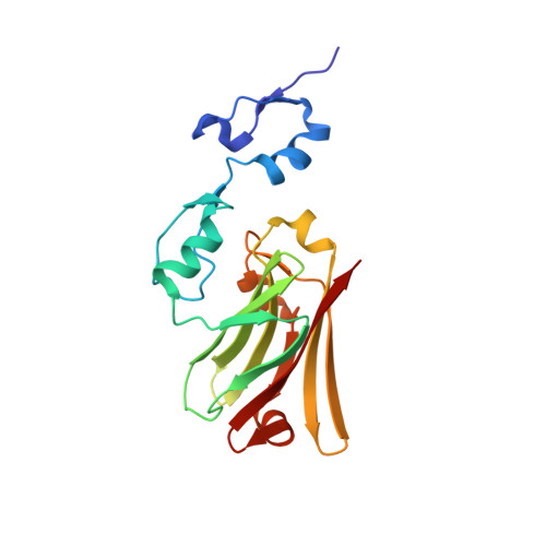
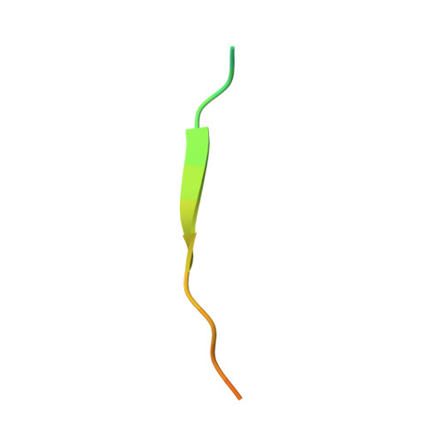
Elucidation of the substrate binding site of Siah ubiquitin ligase

House, C.M.Hancock, N.C.Moller, A.Cromer, B.A.Fedorov, V.Bowtell, D.D.L.Parker, M.W.Polekhina, G.

(2006) Structure 14: 695-701

  • DOI: https://doi.org/10.1016/j.str.2005.12.013
  • Primary Citation of Related Structures:  
    2AN6

  • PubMed Abstract: 

    The Siah family of RING proteins function as ubiquitin ligase components, contributing to the degradation of multiple targets involved in cell growth, differentiation, angiogenesis, oncogenesis, and inflammation. Previously, a binding motif (degron) was recognized in many of the Siah degradation targets, suggesting that Siah itself may facilitate substrate recognition. We report the crystal structure of the Siah in complex with a peptide containing the degron motif. Binding is within a groove formed in part by the zinc fingers and the first two beta strands of the TRAF-C domain of Siah. We show that residues in the degron, previously described to facilitate binding to Siah, interact with the protein. Mutagenesis of Siah at sites of interaction also abrogates both in vitro peptide binding and destabilization of a known Siah target.


  • Organizational Affiliation

    Ian Potter Foundation Centre for Cancer Genomics and Predictive Medicine, Peter MacCallum Cancer Centre, Locked Bag 1 A'Beckett Street, Melbourne, Victoria 8006, Australia.


Macromolecules
Find similar proteins by:  (by identity cutoff)  |  3D Structure
Entity ID: 1
MoleculeChains Sequence LengthOrganismDetailsImage
Ubiquitin ligase SIAH1A
A, B, C, D
191Mus musculusMutation(s): 0 
EC: 6.3.2
UniProt
Find proteins for P61092 (Mus musculus)
Explore P61092 
Go to UniProtKB:  P61092
Entity Groups  
Sequence Clusters30% Identity50% Identity70% Identity90% Identity95% Identity100% Identity
UniProt GroupP61092
Sequence Annotations
Expand
  • Reference Sequence

Find similar proteins by:  Sequence   |   3D Structure  

Entity ID: 2
MoleculeChains Sequence LengthOrganismDetailsImage
peptide from Phyllopod
E, F, G, H
24N/AMutation(s): 0 
Entity Groups  
Sequence Clusters30% Identity50% Identity70% Identity90% Identity95% Identity100% Identity
Sequence Annotations
Expand
  • Reference Sequence
Experimental Data & Validation

Experimental Data

  • Method: X-RAY DIFFRACTION
  • Resolution: 3.00 Å
  • R-Value Free: 0.305 
  • R-Value Work: 0.237 
  • R-Value Observed: 0.241 
  • Space Group: P 1 21 1
Unit Cell:
Length ( Å )Angle ( ˚ )
a = 63.3α = 90
b = 100β = 104.3
c = 103.4γ = 90
Software Package:
Software NamePurpose
REFMACrefinement
DENZOdata reduction
SCALEPACKdata scaling
MOLREPphasing

Structure Validation

View Full Validation Report


Entry History 

Deposition Data

Revision History  (Full details and data files)

  • Version 1.0: 2006-05-30
    Type: Initial release
  • Version 1.1: 2008-04-30
    Changes: Version format compliance
  • Version 1.2: 2011-07-13
    Changes: Version format compliance
  • Version 1.3: 2024-03-13
    Changes: Data collection, Database references, Derived calculations, Refinement description