Crystal structure of a maltotetraose-forming exo-amylase from Pseudomonas stutzeri.
Morishita, Y., Hasegawa, K., Matsuura, Y., Katsube, Y., Kubota, M., Sakai, S.(1997) J Mol Biology 267: 661-672
- PubMed: 9126844 Search on PubMed
- DOI: https://doi.org/10.1006/jmbi.1996.0887
- Primary Citation Related Structures:
2AMG - PubMed Abstract:
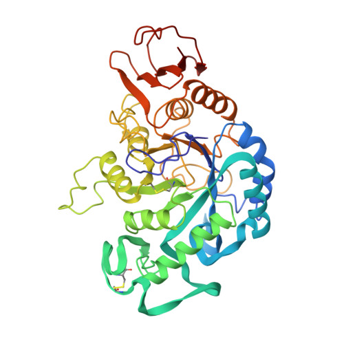
The three-dimensional structure of an exo-type alpha-amylase from Pseudomonas stutzeri which degrades starch from its non-reducing end to produce maltotetraose has been determined by X-ray structure analysis. The catalytic domain of this enzyme (G4-2), whose structure was determined, is a product of spontaneous limited proteolysis in culture broth. It has 429 amino acid residues and a molecular mass of 47,200, and crystallizes in ammonium sulfate solution at pH 7.5. The structure was elucidated by the multiple isomorphous replacement method and refined at 2.0 A resolution, resulting in a final R-factor of 0.178 for significant reflections with a root-mean-square deviation from ideality in bond distances of 0.013 A. The polypeptide chain of this molecule folds into three domains; the first with a (beta/alpha)8 barrel structure, the second with an excursed part from the first one, and the third with five-stranded antiparallel beta-sheets. The active cleft is formed on the C-terminal side of the beta-sheets in the (beta/alpha)8 barrel as in the known endo-type alpha-amylases. A histidine side-chain nitrogen ND1 is coordinated to one of the bound calcium ion. The recognition site of the non-reducing end of the amylose that determines exo-wise degradation is presumed to be at one end of this cleft where there is a disordered loop consisting of the 66th to 72nd residues, and a loop carrying an aspartic acid (Asp160). These structural features may be responsible for the binding of the non-reducing end of the substrate amylose.
- Institute for Protein Research, Osaka University, Suita, Japan.
Organizational Affiliation: