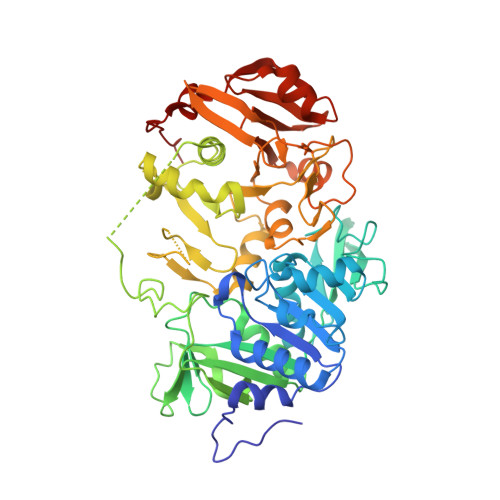
2AHV

Crystal Structure of Acyl-CoA transferase from E. coli O157:H7 (YdiF)-thioester complex with CoA- 1


Experimental Data Snapshot

  • Method: X-RAY DIFFRACTION
  • Resolution: 2.00 Å
  • R-Value Free: 0.224 
  • R-Value Work: 0.182 
  • R-Value Observed: 0.186 

Starting Model: experimental
View more details

wwPDB Validation   3D Report Full Report


Ligand Structure Quality Assessment 


This is version 1.3 of the entry. See complete history


Literature

Crystallographic trapping of the glutamyl-CoA thioester intermediate of family I CoA transferases.

Rangarajan, E.S.Li, Y.Ajamian, E.Iannuzzi, P.Kernaghan, S.D.Fraser, M.E.Cygler, M.Matte, A.

(2005) J Biol Chem 280: 42919-42928

  • DOI: https://doi.org/10.1074/jbc.M510522200
  • Primary Citation of Related Structures:  
    2AHU, 2AHV, 2AHW

  • PubMed Abstract: 

    Coenzyme A transferases are involved in a broad range of biochemical processes in both prokaryotes and eukaryotes, and exhibit a diverse range of substrate specificities. The YdiF protein from Escherichia coli O157:H7 is an acyl-CoA transferase of unknown physiological function, and belongs to a large sequence family of CoA transferases, present in bacteria to humans, which utilize oxoacids as acceptors. In vitro measurements showed that YdiF displays enzymatic activity with short-chain acyl-CoAs. The crystal structures of YdiF and its complex with CoA, the first co-crystal structure for any Family I CoA transferase, have been determined and refined at 1.9 and 2.0 A resolution, respectively. YdiF is organized into tetramers, with each monomer having an open alpha/beta structure characteristic of Family I CoA transferases. Co-crystallization of YdiF with a variety of CoA thioesters in the absence of acceptor carboxylic acid resulted in trapping a covalent gamma-glutamyl-CoA thioester intermediate. The CoA binds within a well defined pocket at the N- and C-terminal domain interface, but makes contact only with the C-terminal domain. The structure of the YdiF complex provides a basis for understanding the different catalytic steps in the reaction of Family I CoA transferases.


  • Organizational Affiliation

    Department of Biochemistry, McGill University, Quebec, Canada.


Macromolecules
Find similar proteins by:  (by identity cutoff)  |  3D Structure
Entity ID: 1
MoleculeChains Sequence LengthOrganismDetailsImage
putative enzyme YdiF
A, B, C, D
531Escherichia coli O157:H7Mutation(s): 0 
Gene Names: YdiF
EC: 2.8.3
UniProt
Find proteins for Q8X5X6 (Escherichia coli O157:H7)
Explore Q8X5X6 
Go to UniProtKB:  Q8X5X6
Entity Groups  
Sequence Clusters30% Identity50% Identity70% Identity90% Identity95% Identity100% Identity
UniProt GroupQ8X5X6
Sequence Annotations
Expand
  • Reference Sequence
Experimental Data & Validation

Experimental Data

  • Method: X-RAY DIFFRACTION
  • Resolution: 2.00 Å
  • R-Value Free: 0.224 
  • R-Value Work: 0.182 
  • R-Value Observed: 0.186 
  • Space Group: P 1 21 1
Unit Cell:
Length ( Å )Angle ( ˚ )
a = 81.085α = 90
b = 140.24β = 108.22
c = 112.682γ = 90
Software Package:
Software NamePurpose
REFMACrefinement
HKL-2000data reduction
SCALEPACKdata scaling
MOLREPphasing

Structure Validation

View Full Validation Report


Ligand Structure Quality Assessment 


Entry History 

Revision History  (Full details and data files)

  • Version 1.0: 2005-11-01
    Type: Initial release
  • Version 1.1: 2008-04-30
    Changes: Version format compliance
  • Version 1.2: 2011-07-13
    Changes: Source and taxonomy, Version format compliance
  • Version 1.3: 2023-08-23
    Changes: Data collection, Database references, Derived calculations, Refinement description