2AGK

Structure of S. cerevisiae His6 protein


Experimental Data Snapshot

  • Method: X-RAY DIFFRACTION
  • Resolution: 1.30 Å
  • R-Value Free: 0.216 
  • R-Value Work: 0.191 

wwPDB Validation   3D Report Full Report


This is version 1.4 of the entry. See complete history


Literature

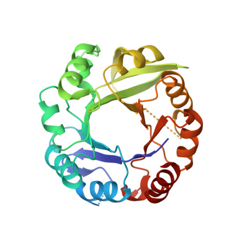
Crystal structure of the yeast His6 enzyme suggests a reaction mechanism

Quevillon-Cheruel, S.Leulliot, N.Graille, M.Blondeau, K.Janin, J.Tilbeurgh, H.V.

(2006) Protein Sci 15: 1516-1521

  • DOI: https://doi.org/10.1110/ps.062144406
  • Primary Citation of Related Structures:  
    2AGK

  • PubMed Abstract: 

    The Saccharomyces cerevisiae His6 gene codes for the enzyme phosphoribosyl-5-amino-1-phosphoribosyl-4-imidazolecarboxamide isomerase, catalyzing the fourth step in histidine biosynthesis. To get an insight into the structure and function of this enzyme, we determined its X-ray structure at a resolution of 1.30 A using the anomalous diffraction signal of the protein's sulphur atoms at 1.77 A wavelength. His6 folds in an (alpha/beta)8 barrel similar to HisA, which performs the same function in bacteria and archaea. We found a citrate molecule from the buffer bound in a pocket near the expected position of the active site and used it to model the open form of the substrate (phosphoribulosyl moiety), which is a reaction intermediate. This model enables us to identify catalytic residues and to propose a reaction mechanism where two aspartates act as acid/base catalysts: Asp134 as a proton donor for ring opening, and Asp9 as a proton acceptor and donor during enolization of the aminoaldose. Asp9 is conserved in yeast His6 and bacterial or archaeal HisA sequences, and Asp134 has equivalents in both HisA and TrpF, but they occur at a different position in the protein sequence.


  • Organizational Affiliation

    Institut de Biochimie et de Biophysique Moléculaire et Cellulaire (CNRS-UMR 8619), Université Paris-Sud, 91405 Orsay, France.


Macromolecules
Find similar proteins by:  (by identity cutoff)  |  3D Structure
Entity ID: 1
MoleculeChains Sequence LengthOrganismDetailsImage
1-(5-phosphoribosyl)-5-[(5-phosphoribosylamino)methylideneamino] imidazole-4-carboxamide isomerase260Saccharomyces cerevisiaeMutation(s): 0 
Gene Names: YIL020c
EC: 5.3.1.16
UniProt
Find proteins for P40545 (Saccharomyces cerevisiae (strain ATCC 204508 / S288c))
Explore P40545 
Go to UniProtKB:  P40545
Entity Groups  
Sequence Clusters30% Identity50% Identity70% Identity90% Identity95% Identity100% Identity
UniProt GroupP40545
Sequence Annotations
Expand
  • Reference Sequence
Experimental Data & Validation

Experimental Data

  • Method: X-RAY DIFFRACTION
  • Resolution: 1.30 Å
  • R-Value Free: 0.216 
  • R-Value Work: 0.191 
  • Space Group: C 1 2 1
Unit Cell:
Length ( Å )Angle ( ˚ )
a = 101.863α = 90
b = 71.427β = 104.71
c = 40.57γ = 90
Software Package:
Software NamePurpose
XSCALEdata scaling
REFMACrefinement
PDB_EXTRACTdata extraction
XDSdata reduction
SHARPphasing

Structure Validation

View Full Validation Report


Entry History 

Deposition Data

Revision History  (Full details and data files)

  • Version 1.0: 2006-06-20
    Type: Initial release
  • Version 1.1: 2008-04-01
    Changes: Version format compliance
  • Version 1.2: 2011-07-13
    Changes: Version format compliance
  • Version 1.3: 2017-10-11
    Changes: Refinement description
  • Version 1.4: 2024-03-13
    Changes: Data collection, Database references, Derived calculations