2ZYV | pdb_00002zyv

Crystal structure of mouse cytosolic sulfotransferase mSULT1D1 complex with PAPS/PAP and p-nitrophenol


Experimental Data Snapshot

  • Method: X-RAY DIFFRACTION
  • Resolution: 1.81 Å
  • R-Value Free: 
    0.203 (Depositor), 0.202 (DCC) 
  • R-Value Work: 
    0.179 (Depositor), 0.178 (DCC) 
  • R-Value Observed: 
    0.180 (Depositor) 

Starting Model: experimental
View more details

wwPDB Validation 3D Report Full Report

Validation slider image for 2ZYV

This is version 1.2 of the entry. See complete history

Literature

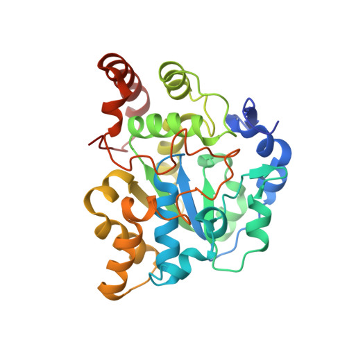
Snapshot of a Michaelis complex in a sulfuryl transfer reaction: Crystal structure of a mouse sulfotransferase, mSULT1D1, complexed with donor substrate and accepter substrate

Teramoto, T.Sakakibara, Y.Liu, M.-C.Suiko, M.Kimura, M.Kakuta, Y.

(2009) Biochem Biophys Res Commun 383: 83-87

  • DOI: https://doi.org/10.1016/j.bbrc.2009.03.146
  • Primary Citation Related Structures: 
    2ZYT, 2ZYU, 2ZYV, 2ZYW

  • PubMed Abstract: 

    We report the crystal structure of mouse sulfotransferase, mSULT1D1, complexed with donor substrate 3'-phosphoadenosine 5'-phosphosulfate and accepter substrate p-nitrophenol. The structure is the first report of the native Michaelis complex of sulfotransferase. In the structure, three proposed catalytic residues (Lys48, Lys106, and His108) were in proper positions for engaging in the sulfuryl transfer reaction. The data strongly support that the sulfuryl transfer reaction proceeds through an S(N)2-like in-line displacement mechanism.


  • Organizational Affiliation
    • Department of Systems Life Sciences, Kyushu University, Hakozaki, Higashi-ku, Fukuoka, Japan.

Macromolecule Content 

  • Total Structure Weight: 36.47 kDa 
  • Atom Count: 2,778 
  • Modeled Residue Count: 289 
  • Deposited Residue Count: 295 
  • Unique protein chains: 1

Macromolecules

Find similar proteins by:|  3D Structure
Entity ID: 1
MoleculeChains  Sequence LengthOrganismDetailsImage
Tyrosine-ester sulfotransferaseA [auth X]295Mus musculusMutation(s): 0 
Gene Names: Sult1d1
EC: 2.8.2.9 (PDB Primary Data), 2.8.2 (UniProt)
UniProt
Find proteins for Q3UZZ6 (Mus musculus)
Explore Q3UZZ6 
Go to UniProtKB:  Q3UZZ6
Entity Groups
Sequence Clusters30% Identity50% Identity70% Identity90% Identity95% Identity100% Identity
UniProt GroupQ3UZZ6
Sequence Annotations
Expand
Reference Sequence

Small Molecules

Ligands 3 Unique
IDChains Name / Formula / InChI Key2D Diagram3D Interactions
PPS

Query on PPS


Download:Ideal Coordinates CCD File
B [auth X]3'-PHOSPHATE-ADENOSINE-5'-PHOSPHATE SULFATE
C10 H15 N5 O13 P2 S
GACDQMDRPRGCTN-KQYNXXCUSA-N
NPO

Query on NPO


Download:Ideal Coordinates CCD File
C [auth X],
D [auth X]
P-NITROPHENOL
C6 H5 N O3
BTJIUGUIPKRLHP-UHFFFAOYSA-N
GOL

Query on GOL


Download:Ideal Coordinates CCD File
E [auth X]
F [auth X]
G [auth X]
H [auth X]
I [auth X]
E [auth X],
F [auth X],
G [auth X],
H [auth X],
I [auth X],
J [auth X]
GLYCEROL
C3 H8 O3
PEDCQBHIVMGVHV-UHFFFAOYSA-N

Experimental Data & Validation

Experimental Data

  • Method: X-RAY DIFFRACTION
  • Resolution: 1.81 Å
  • R-Value Free:  0.203 (Depositor), 0.202 (DCC) 
  • R-Value Work:  0.179 (Depositor), 0.178 (DCC) 
  • R-Value Observed: 0.180 (Depositor) 
Space Group: C 1 2 1
Unit Cell:
Length ( Å )Angle ( ˚ )
a = 155.241α = 90
b = 67.793β = 105.23
c = 42.904γ = 90
Software Package:
Software NamePurpose
REFMACrefinement
BBSdata collection
HKL-2000data reduction
HKL-2000data scaling
MOLREPphasing

Structure Validation

View Full Validation Report


Entry History 

Deposition Data

Revision History  (Full details and data files)

  • Version 1.0: 2009-04-21
    Type: Initial release
  • Version 1.1: 2011-07-13
    Changes: Non-polymer description, Version format compliance
  • Version 1.2: 2023-11-01
    Changes: Data collection, Database references, Derived calculations, Refinement description