Structure of the apo decarbamylated form of 2,3-diketo-5-methylthiopentyl-1-phosphate enolase from Bacillus subtilis
Tamura, H., Saito, Y., Ashida, H., Kai, Y., Inoue, T., Yokota, A., Matsumura, H.(2009) Acta Crystallogr D Biol Crystallogr 65: 942-951
- PubMed: 19690372 Search on PubMed
- DOI: https://doi.org/10.1107/S0907444909022422
- Primary Citation Related Structures:
2ZVI - PubMed Abstract:
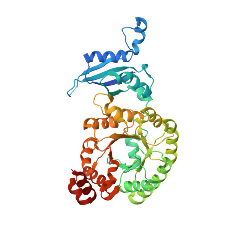
2,3-Diketo-5-methylthiopentyl-1-phosphate enolase (DK-MTP-1P enolase), a RuBisCO-like protein (RLP), catalyzes the enolization of 2,3-diketo-5-methylthiopentyl-1-phosphate. The crystal structure of the apo decarbamylated form (E form) of Bacillus subtilis DK-MTP-1P enolase (Bs-DK-MTP-1P enolase) has been determined at 2.3 A resolution. The overall structure of the E form of Bs-DK-MTP-1P enolase highly resembles that of Geobacillus kaustophilus DK-MTP-1P enolase (Gk-DK-MTP-1P enolase), with the exception of a few insertions or deletions and of a few residues at the active site. In the E form of Bs-DK-MTP-1P enolase, Lys150 (equivalent to Lys175 in RuBisCO) at the active site adopts a conformation that is distinct from those observed in the other forms of Gk-DK-MTP-1P enolase. This unusual conformational change appears to be induced by changes in the varphi and psi angles of Gly151, which is conserved in the sequences of the Bs-DK-MTP-1P and Gk-DK-MTP-1P enolases but not in those of RuBisCOs. The loop at 303-312, equivalent to the catalytic loop termed ;loop-6' in RuBisCO, is in a closed conformation in the E form of Bs-DK-MTP-1P enolase. The closed conformation appears to be stabilized by Pro312, which is conserved in the sequences of several RLPs (equivalent to Glu338 in RuBisCO). Based on these results, the characteristic structural features of DK-MTP-1P enolase are discussed.
- Department of Applied Chemistry, Graduate School of Engineering, Osaka University, Suita, Osaka, Japan.
Organizational Affiliation: