2ZS9 | pdb_00002zs9

Pantothenate kinase from Mycobacterium tuberculosis (MtPanK) in complex with ADP and Pantothenate


Experimental Data Snapshot

  • Method: X-RAY DIFFRACTION
  • Resolution: 2.70 Å
  • R-Value Free: 
    0.233 (Depositor), 0.240 (DCC) 
  • R-Value Work: 
    0.203 (Depositor), 0.204 (DCC) 
  • R-Value Observed: 
    0.203 (Depositor) 

Starting Model: experimental
View more details

wwPDB Validation 3D Report Full Report

Validation slider image for 2ZS9

Ligand Structure Quality Assessment 


This is version 1.2 of the entry. See complete history

Literature

Mycobacterium tuberculosis pantothenate kinase: possible changes in location of ligands during enzyme action

Chetnani, B.Das, S.Kumar, P.Surolia, A.Vijayan, M.

(2009) Acta Crystallogr D Biol Crystallogr 65: 312-325

  • DOI: https://doi.org/10.1107/S0907444909002170
  • Primary Citation Related Structures: 
    2ZS7, 2ZS8, 2ZS9, 2ZSA, 2ZSB, 2ZSD, 2ZSE, 2ZSF

  • PubMed Abstract: 

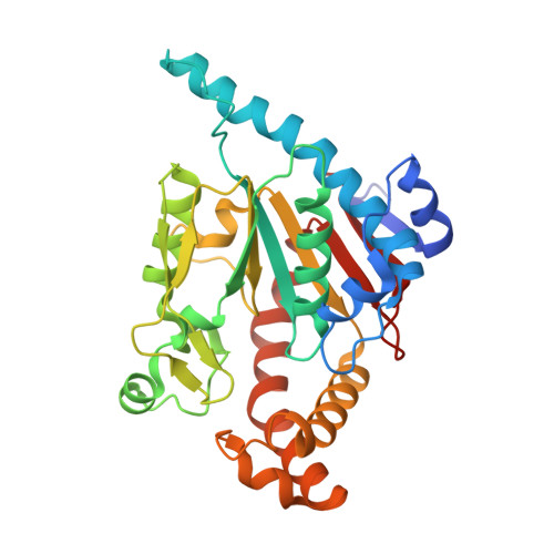
    The crystal structures of complexes of Mycobacterium tuberculosis pantothenate kinase with the following ligands have been determined: (i) citrate; (ii) the nonhydrolysable ATP analogue AMPPCP and pantothenate (the initiation complex); (iii) ADP and phosphopantothenate resulting from phosphorylation of pantothenate by ATP in the crystal (the end complex); (iv) ATP and ADP, each with half occupancy, resulting from a quick soak of crystals in ATP (the intermediate complex); (v) CoA; (vi) ADP prepared by soaking and cocrystallization, which turned out to have identical structures, and (vii) ADP and pantothenate. Solution studies on CoA binding and catalytic activity have also been carried out. Unlike in the case of the homologous Escherichia coli enzyme, AMPPCP and ADP occupy different, though overlapping, locations in the respective complexes; the same is true of pantothenate in the initiation complex and phosphopantothenate in the end complex. The binding site of MtPanK is substantially preformed, while that of EcPanK exhibits considerable plasticity. The difference in the behaviour of the E. coli and M. tuberculosis enzymes could be explained in terms of changes in local structure resulting from substitutions. It is unusual for two homologous enzymes to exhibit such striking differences in action. Therefore, the results have to be treated with caution. However, the changes in the locations of ligands exhibited by M. tuberculosis pantothenate kinase are remarkable and novel.


  • Organizational Affiliation
    • Molecular Biophysics Unit, Indian Institute of Science, Bangalore, India.

Macromolecule Content 

  • Total Structure Weight: 37.16 kDa 
  • Atom Count: 2,757 
  • Modeled Residue Count: 307 
  • Deposited Residue Count: 312 
  • Unique protein chains: 1

Macromolecules

Find similar proteins by:|  3D Structure
Entity ID: 1
MoleculeChains  Sequence LengthOrganismDetailsImage
Pantothenate kinase312Mycobacterium tuberculosisMutation(s): 0 
Gene Names: coaA (Rv1092c)
EC: 2.7.1.33
UniProt
Find proteins for P9WPA7 (Mycobacterium tuberculosis (strain ATCC 25618 / H37Rv))
Explore P9WPA7 
Go to UniProtKB:  P9WPA7
Entity Groups
Sequence Clusters30% Identity50% Identity70% Identity90% Identity95% Identity100% Identity
UniProt GroupP9WPA7
Sequence Annotations
Expand
Reference Sequence

Small Molecules

Ligands 4 Unique
IDChains Name / Formula / InChI Key2D Diagram3D Interactions
ADP

Query on ADP


Download:Ideal Coordinates CCD File
B [auth A]ADENOSINE-5'-DIPHOSPHATE
C10 H15 N5 O10 P2
XTWYTFMLZFPYCI-KQYNXXCUSA-N
PAU

Query on PAU


Download:Ideal Coordinates CCD File
N [auth A]PANTOTHENOIC ACID
C9 H17 N O5
GHOKWGTUZJEAQD-ZETCQYMHSA-N
GOL

Query on GOL


Download:Ideal Coordinates CCD File
C [auth A]
D [auth A]
E [auth A]
F [auth A]
G [auth A]
C [auth A],
D [auth A],
E [auth A],
F [auth A],
G [auth A],
H [auth A],
I [auth A],
J [auth A]
GLYCEROL
C3 H8 O3
PEDCQBHIVMGVHV-UHFFFAOYSA-N
NA

Query on NA


Download:Ideal Coordinates CCD File
K [auth A],
L [auth A],
M [auth A]
SODIUM ION
Na
FKNQFGJONOIPTF-UHFFFAOYSA-N

Experimental Data & Validation

Experimental Data

  • Method: X-RAY DIFFRACTION
  • Resolution: 2.70 Å
  • R-Value Free:  0.233 (Depositor), 0.240 (DCC) 
  • R-Value Work:  0.203 (Depositor), 0.204 (DCC) 
  • R-Value Observed: 0.203 (Depositor) 
Space Group: P 31 2 1
Unit Cell:
Length ( Å )Angle ( ˚ )
a = 104.194α = 90
b = 104.194β = 90
c = 90.304γ = 120
Software Package:
Software NamePurpose
CNSrefinement
MAR345dtbdata collection
DENZOdata reduction
SCALEPACKdata scaling
AMoREphasing

Structure Validation

View Full Validation Report


Ligand Structure Quality Assessment 


Entry History 

Deposition Data

Revision History  (Full details and data files)

  • Version 1.0: 2009-07-21
    Type: Initial release
  • Version 1.1: 2011-07-13
    Changes: Non-polymer description, Version format compliance
  • Version 1.2: 2023-11-01
    Changes: Data collection, Database references, Derived calculations, Refinement description