Crystal Structure of the HA3 Subcomponent of Clostridium botulinum Type C Progenitor Toxin
Nakamura, T., Kotani, M., Tonozuka, T., Ide, A., Oguma, K., Nishikawa, A.(2009) J Mol Biology 385: 1193-1206
- PubMed: 19071137 Search on PubMed
- DOI: https://doi.org/10.1016/j.jmb.2008.11.039
- Primary Citation Related Structures:
2ZOE, 2ZS6 - PubMed Abstract:
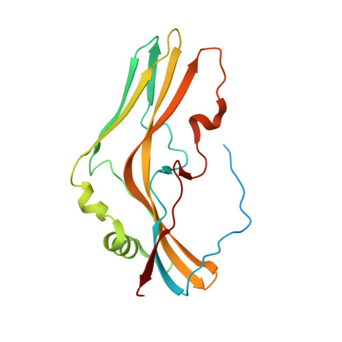
The Clostridium botulinum type C 16S progenitor toxin contains a neurotoxin and several nontoxic components, designated nontoxic nonhemagglutinin (HA), HA1 (HA-33), HA2 (HA-17), HA3a (HA-22-23), and HA3b (HA-53). The HA3b subcomponent seems to play an important role cooperatively with HA1 in the internalization of the toxin by gastrointestinal epithelial cells via binding of these subcomponents to specific oligosaccharides. In this study, we investigated the sugar-binding specificity of the HA3b subcomponent using recombinant protein fused to glutathione S-transferase and determined the three-dimensional structure of the HA3a-HA3b complex based on X-ray crystallography. The crystal structure was determined at a resolution of 2.6 A. HA3b contains three domains, domains I to III, and the structure of domain I resembles HA3a. In crystal packing, three HA3a-HA3b molecules are assembled to form a three-leaved propeller-like structure. The three HA3b domain I and three HA3a alternate, forming a trimer of dimers. In a database search, no proteins with high structural homology to any of the domains (Z score >10) were found. Especially, HA3a and HA3b domain I, mainly composed of beta-sheets, reveal a unique fold. In binding assays, HA3b bound sialic acid with high affinity, but did not bind galactose, N-acetylgalactosamine, or N-acetylglucosamine. The electron density of liganded N-acetylneuraminic acid was determined by crystal soaking. In the sugar-complex structure, the N-acetylneuraminic acid-binding site was located in the cleft formed between domains II and III of HA3b. This report provides the first determination of the three-dimensional structure of the HA3a-HA3b complex and its sialic acid binding site. Our results will provide useful information for elucidating the mechanism of assembly of the C16S toxin and for understanding the interactions with oligosaccharides on epithelial cells and internalization of the botulinum toxin complex.
- Department of Applied Biological Science, Tokyo University of Agriculture and Technology, 3-5-8 Saiwai-cho, Fuchu-shi, Tokyo 183-8509, Japan.
Organizational Affiliation: