2ZPT | pdb_00002zpt

Crystal structure of mouse sulfotransferase SULT1D1 complex with PAP


Experimental Data Snapshot

  • Method: X-RAY DIFFRACTION
  • Resolution: 1.15 Å
  • R-Value Free: 
    0.160 (Depositor), 0.160 (DCC) 
  • R-Value Work: 
    0.148 (Depositor), 0.147 (DCC) 
  • R-Value Observed: 
    0.149 (Depositor) 

Starting Model: experimental
View more details

wwPDB Validation 3D Report Full Report

Validation slider image for 2ZPT

Ligand Structure Quality Assessment 


This is version 1.2 of the entry. See complete history

Literature

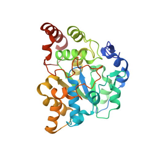
Crystal structure of mSULT1D1, a mouse catecholamine sulfotransferase

Teramoto, T.Sakakibara, Y.Inada, K.Kurogi, K.Liu, M.-C.Suiko, M.Kimura, M.Kakuta, Y.

(2008) FEBS Lett 582: 3909-3914

  • DOI: https://doi.org/10.1016/j.febslet.2008.10.035
  • Primary Citation Related Structures: 
    2ZPT

  • PubMed Abstract: 

    In mammals, sulfonation as mediated by specific cytosolic sulfotransferases (SULTs) plays an important role in the homeostasis of dopamine and other catecholamines. To gain insight into the structural basis for dopamine recognition/binding, we determined the crystal structure of a mouse dopamine-sulfating SULT, mouse SULT1D1 (mSULT1D1). Data obtained indicated that mSULT1D1 comprises of a single alpha/beta domain with a five-stranded parallel beta-sheet. In contrast to the structure of the human SULT1A3 (hSULT1A3)-dopamine complex previously reported, molecular modeling and mutational analysis revealed that a water molecule plays a critical role in the recognition of the amine group of dopamine by mSULT1D1. These results imply differences in substrate binding between dopamine-sulfating SULTs from different species.


  • Organizational Affiliation
    • Laboratory of Structural Biology, Department of Systems Life Sciences, Japan.

Macromolecule Content 

  • Total Structure Weight: 35.65 kDa 
  • Atom Count: 3,162 
  • Modeled Residue Count: 288 
  • Deposited Residue Count: 295 
  • Unique protein chains: 1

Macromolecules

Find similar proteins by:|  3D Structure
Entity ID: 1
MoleculeChains  Sequence LengthOrganismDetailsImage
Tyrosine-ester sulfotransferaseA [auth X]295Mus musculusMutation(s): 0 
Gene Names: Sult1d1
EC: 2.8.2.9 (PDB Primary Data), 2.8.2 (UniProt)
UniProt
Find proteins for Q3UZZ6 (Mus musculus)
Explore Q3UZZ6 
Go to UniProtKB:  Q3UZZ6
Entity Groups
Sequence Clusters30% Identity50% Identity70% Identity90% Identity95% Identity100% Identity
UniProt GroupQ3UZZ6
Sequence Annotations
Expand
Reference Sequence

Small Molecules

Experimental Data & Validation

Experimental Data

  • Method: X-RAY DIFFRACTION
  • Resolution: 1.15 Å
  • R-Value Free:  0.160 (Depositor), 0.160 (DCC) 
  • R-Value Work:  0.148 (Depositor), 0.147 (DCC) 
  • R-Value Observed: 0.149 (Depositor) 
Space Group: C 1 2 1
Unit Cell:
Length ( Å )Angle ( ˚ )
a = 155.819α = 90
b = 67.6β = 105.63
c = 42.696γ = 90
Software Package:
Software NamePurpose
REFMACrefinement
BBSdata collection
HKL-2000data reduction
HKL-2000data scaling
MOLREPphasing

Structure Validation

View Full Validation Report


Ligand Structure Quality Assessment 


Entry History 

Deposition Data

Revision History  (Full details and data files)

  • Version 1.0: 2008-11-18
    Type: Initial release
  • Version 1.1: 2011-07-13
    Changes: Non-polymer description, Version format compliance
  • Version 1.2: 2023-11-01
    Changes: Data collection, Database references, Derived calculations, Refinement description