2ZGD | pdb_00002zgd

Asn-hydroxylation stabilises the ankyrin repeat domain fold


Experimental Data Snapshot

  • Method: X-RAY DIFFRACTION
  • Resolution: 1.90 Å
  • R-Value Free: 
    0.224 (Depositor), 0.225 (DCC) 
  • R-Value Work: 
    0.173 (Depositor), 0.178 (DCC) 

Starting Model: experimental
View more details

wwPDB Validation 3D Report Full Report

Validation slider image for 2ZGD

This is version 1.3 of the entry. See complete history

Literature

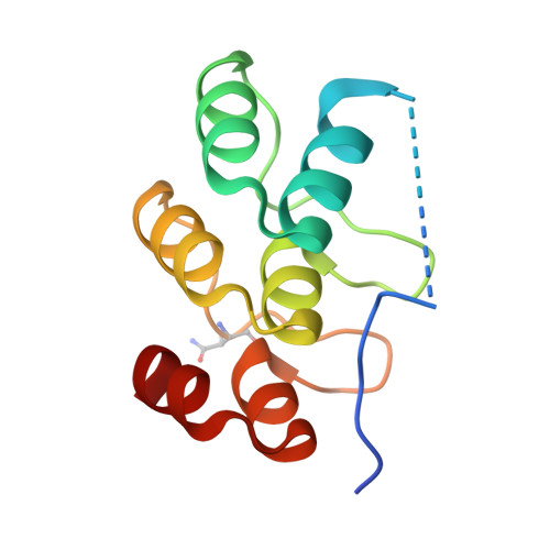
Asparagine beta-hydroxylation stabilizes the ankyrin repeat domain fold

Kelly, L.McDonough, M.A.Coleman, M.L.Ratcliffe, P.J.Schofield, C.J.

(2009) Mol Biosyst 5: 52-58

  • DOI: https://doi.org/10.1039/b815271c
  • Primary Citation Related Structures: 
    2ZGD, 2ZGG

  • PubMed Abstract: 

    Ankyrin repeats (ARs) are one of the most common structural motifs among eukaryotic proteins. Recent analyses have shown that factor inhibiting hypoxia-inducible factor (FIH) catalyses the hydroxylation of highly conserved Asn-residues within ankyrin repeat domains (ARDs). However, the effect of Asn-hydroxylation on ARD structure is unknown. Supporting the proposal that FIH-mediated ARD hydroxylation is ubiquitous we report that consensus ARD proteins are FIH substrates both in vitro and in vivo. X-ray diffraction analyses revealed that hydroxylation does not alter the archetypical ARD conformation in the crystalline state. However, other biophysical analyses revealed that hydroxylation significantly stabilizes the ARD fold in solution. We propose that intracellular protein hydroxylation is much more common than previously thought and that one of its roles is stabilization of localized regions of ARD folds.


  • Organizational Affiliation
    • The Chemistry Research Laboratory and Oxford Centre for Integrative Systems Biology, University of Oxford, Oxford, UKOX1 3TA.

Macromolecule Content 

  • Total Structure Weight: 12.29 kDa 
  • Atom Count: 782 
  • Modeled Residue Count: 97 
  • Deposited Residue Count: 110 
  • Unique protein chains: 1

Macromolecules

Find similar proteins by:|  3D Structure
Entity ID: 1
MoleculeChains  Sequence LengthOrganismDetailsImage
3 repeat synthetic ankyrin110N/AMutation(s): 0 

Small Molecules

Experimental Data & Validation

Experimental Data

  • Method: X-RAY DIFFRACTION
  • Resolution: 1.90 Å
  • R-Value Free:  0.224 (Depositor), 0.225 (DCC) 
  • R-Value Work:  0.173 (Depositor), 0.178 (DCC) 
Space Group: P 31 2 1
Unit Cell:
Length ( Å )Angle ( ˚ )
a = 48.887α = 90
b = 48.887β = 90
c = 67.088γ = 120
Software Package:
Software NamePurpose
DENZOdata reduction
SCALEPACKdata scaling
PHASERphasing
CNSrefinement
PDB_EXTRACTdata extraction
HKL-2000data collection
HKL-2000data reduction
HKL-2000data scaling

Structure Validation

View Full Validation Report


Entry History 

Deposition Data

Revision History  (Full details and data files)

  • Version 1.0: 2008-02-05
    Type: Initial release
  • Version 1.1: 2011-07-13
    Changes: Version format compliance
  • Version 1.2: 2017-10-11
    Changes: Refinement description
  • Version 1.3: 2023-11-01
    Changes: Data collection, Database references, Derived calculations, Refinement description