2ZFX

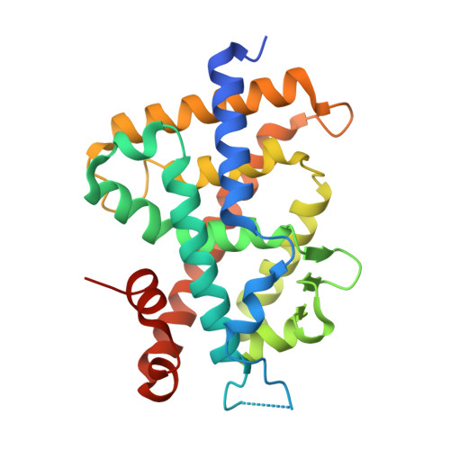
Crystal structure of the rat vitamin D receptor ligand binding domain complexed with YR301 and a synthetic peptide containing the NR2 box of DRIP 205


Experimental Data Snapshot

  • Method: X-RAY DIFFRACTION
  • Resolution: 1.99 Å
  • R-Value Free: 0.258 
  • R-Value Work: 0.206 
  • R-Value Observed: 0.208 

wwPDB Validation   3D Report Full Report


Ligand Structure Quality Assessment 


This is version 1.4 of the entry. See complete history


Literature

Structure of the ligand-binding domain of rat VDR in complex with the nonsecosteroidal vitamin D3 analogue YR301

Kakuda, S.Okada, K.Eguchi, H.Takenouchi, K.Hakamata, W.Kurihara, M.Takimoto-Kamimura, M.

(2008) Acta Crystallogr Sect F Struct Biol Cryst Commun 64: 970-973

  • DOI: https://doi.org/10.1107/S1744309108026754
  • Primary Citation of Related Structures:  
    2ZFX

  • PubMed Abstract: 

    Vitamin D receptor (VDR) is a ligand-inducible hormone receptor that mediates 1alpha,25(OH)(2)D(3) action, regulating calcium and phosphate metabolism, induces potent cell differentiation activity and has immunosuppressive effects. Analogues of 1alpha,25(OH)(2)D(3) have been used clinically for some years. However, the risk of potential side effects limits the use of these substances. LG190178 is a novel nonsecosteroidal ligand for VDR. (2S)-3-[4-(3-{4-[(2R)-2-hydroxy-3,3-dimethylbutoxy]-3-methylphenyl}pentan-3-yl)-2-methylphenoxy] propane-1,2-diol (YR301) is the only one of the four evaluated stereoisomers of LG190178 to have strong activity. To understand the strong activity of YR301, the crystal structure of YR301 complexed with the rat VDR ligand-binding domain (VDR LBD) was solved at 2.0 A resolution and compared with the structure of the VDR LBD-1alpha,25(OH)(2)D(3) complex. YR301 and 1alpha,25(OH)(2)D(3) share the same position and the diethylmethyl group occupies a similar space to the C and D rings of 1alpha,25(OH)(2)D(3). YR301 has two characteristic hydroxyl groups which contribute to its potent activity. The first is 2'-OH, which forms hydrogen bonds to the NE2 atoms of both His301 and His393. The other is 2-OH, which interacts with Ser233 OG and Arg270 NH1. These two hydroxyl groups of YR301 correspond exactly to 25-OH and 1-OH, respectively, of 1alpha,25(OH)(2)D(3). The terminal hydroxyl group (3-OH) of YR301 is directly hydrogen bonded to Arg270 and also interacts indirectly with Tyr232 OH and the backbone NH of Asp144 via water molecules. Additional derivatization of the terminal hydroxyl group using the positions of the water molecules might be useful for the design of more potent compounds.


  • Organizational Affiliation

    Teijin Institute for Biomedical Research, Japan. s.kakuda@teijin.co.jp


Macromolecules
Find similar proteins by:  (by identity cutoff)  |  3D Structure
Entity ID: 1
MoleculeChains Sequence LengthOrganismDetailsImage
Vitamin D3 receptor265Rattus norvegicusMutation(s): 0 
UniProt
Find proteins for P13053 (Rattus norvegicus)
Explore P13053 
Go to UniProtKB:  P13053
Entity Groups  
Sequence Clusters30% Identity50% Identity70% Identity90% Identity95% Identity100% Identity
UniProt GroupP13053
Sequence Annotations
Expand
  • Reference Sequence

Find similar proteins by:  Sequence   |   3D Structure  

Entity ID: 2
MoleculeChains Sequence LengthOrganismDetailsImage
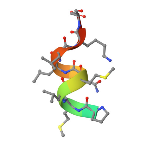
DRIP 205 NR2 box peptideB [auth C]13synthetic constructMutation(s): 0 
UniProt & NIH Common Fund Data Resources
Find proteins for Q15648 (Homo sapiens)
Explore Q15648 
Go to UniProtKB:  Q15648
PHAROS:  Q15648
GTEx:  ENSG00000125686 
Entity Groups  
Sequence Clusters30% Identity50% Identity70% Identity90% Identity95% Identity100% Identity
UniProt GroupQ15648
Sequence Annotations
Expand
  • Reference Sequence
Small Molecules
Ligands 1 Unique
IDChains Name / Formula / InChI Key2D Diagram3D Interactions
YR3
Query on YR3

Download Ideal Coordinates CCD File 
C [auth A](2S)-3-{4-[1-ethyl-1-(4-{[(2R)-2-hydroxy-3,3-dimethylbutyl]oxy}-3-methylphenyl)propyl]-2-methylphenoxy}propane-1,2-diol
C28 H42 O5
AIKLCYAFOOBGEV-OZXSUGGESA-N
Experimental Data & Validation

Experimental Data

  • Method: X-RAY DIFFRACTION
  • Resolution: 1.99 Å
  • R-Value Free: 0.258 
  • R-Value Work: 0.206 
  • R-Value Observed: 0.208 
  • Space Group: P 21 21 2
Unit Cell:
Length ( Å )Angle ( ˚ )
a = 44.188α = 90
b = 136.344β = 90
c = 47.29γ = 90
Software Package:
Software NamePurpose
REFMACrefinement

Structure Validation

View Full Validation Report


Ligand Structure Quality Assessment 


Entry History 

Deposition Data

Revision History  (Full details and data files)

  • Version 1.0: 2009-01-20
    Type: Initial release
  • Version 1.1: 2011-07-13
    Changes: Version format compliance
  • Version 1.2: 2017-08-16
    Changes: Source and taxonomy
  • Version 1.3: 2017-08-23
    Changes: Data collection, Source and taxonomy
  • Version 1.4: 2024-03-13
    Changes: Data collection, Database references, Derived calculations