2ZDS

Crystal Structure of SCO6571 from Streptomyces coelicolor A3(2)


Experimental Data Snapshot

  • Method: X-RAY DIFFRACTION
  • Resolution: 2.30 Å
  • R-Value Free: 0.210 
  • R-Value Work: 0.167 
  • R-Value Observed: 0.167 

wwPDB Validation   3D Report Full Report


This is version 1.3 of the entry. See complete history


Literature

Crystal structure of SCO6571 from Streptomyces coelicolor A3(2).

Begum, P.Sakai, N.Hayashi, T.Gao, Y.G.Tamura, T.Watanabe, N.Yao, M.Tanaka, I.

(2008) Protein Pept Lett 15: 709-712

  • DOI: https://doi.org/10.2174/092986608785133636
  • Primary Citation of Related Structures:  
    2ZDS

  • PubMed Abstract: 

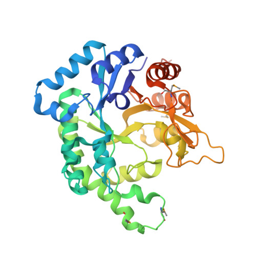
    SCO6571 protein from Streptomyces coelicolor A3(2) was overexpressed and purified using Rhodococcus erythropolis as an expressing host. Crystals of selenomethionine-substituted SCO6571 have been obtained by vapor diffusion method. SCO6571 crystals diffract to 2.3 A and were found to belong to the orthorhombic space group P2(1)2(1)2(1) with unit cell parameters a = 84.5, b = 171.6, c = 184.8 A. Six molecules in the asymmetric unit give a crystal volume per protein mass (V(M)) of 2.97 A (3) Da(-1) and solvent content of 58.6 %. The structure was solved by the single wavelength anomalous diffraction (SAD) method. SCO6571 is a TIM-barrel fold protein that assembles into a hexameric molecule with D(3) symmetry.


  • Organizational Affiliation

    Faculty of Advanced Life Sciences, Hokkaido University, Sapporo 060-0810, Japan.


Macromolecules
Find similar proteins by:  (by identity cutoff)  |  3D Structure
Entity ID: 1
MoleculeChains Sequence LengthOrganismDetailsImage
Putative DNA-binding protein
A, B, C, D, E
A, B, C, D, E, F
340Streptomyces coelicolorMutation(s): 0 
Gene Names: sco6571
UniProt
Find proteins for O69946 (Streptomyces coelicolor (strain ATCC BAA-471 / A3(2) / M145))
Explore O69946 
Go to UniProtKB:  O69946
Entity Groups  
Sequence Clusters30% Identity50% Identity70% Identity90% Identity95% Identity100% Identity
UniProt GroupO69946
Sequence Annotations
Expand
  • Reference Sequence
Small Molecules
Modified Residues  1 Unique
IDChains TypeFormula2D DiagramParent
MSE
Query on MSE
A, B, C, D, E
A, B, C, D, E, F
L-PEPTIDE LINKINGC5 H11 N O2 SeMET
Experimental Data & Validation

Experimental Data

  • Method: X-RAY DIFFRACTION
  • Resolution: 2.30 Å
  • R-Value Free: 0.210 
  • R-Value Work: 0.167 
  • R-Value Observed: 0.167 
  • Space Group: P 21 21 21
Unit Cell:
Length ( Å )Angle ( ˚ )
a = 84.481α = 90
b = 171.593β = 90
c = 184.796γ = 90
Software Package:
Software NamePurpose
HKL-2000data collection
SOLVEphasing
REFMACrefinement
HKL-2000data reduction
HKL-2000data scaling

Structure Validation

View Full Validation Report


Entry History 

Deposition Data

Revision History  (Full details and data files)

  • Version 1.0: 2008-12-02
    Type: Initial release
  • Version 1.1: 2011-07-13
    Changes: Version format compliance
  • Version 1.2: 2017-10-11
    Changes: Refinement description
  • Version 1.3: 2019-10-16
    Changes: Data collection, Database references, Derived calculations