2ZCM | pdb_00002zcm

Crystal structure of IcaR, a repressor of the TetR family


Experimental Data Snapshot

  • Method: X-RAY DIFFRACTION
  • Resolution: 1.33 Å
  • R-Value Free: 
    0.223 (Depositor), 0.218 (DCC) 
  • R-Value Work: 
    0.190 (Depositor), 0.190 (DCC) 

wwPDB Validation 3D Report Full Report

Validation slider image for 2ZCM

This is version 1.3 of the entry. See complete history

Literature

Crystal structure of IcaR, a repressor of the TetR family implicated in biofilm formation in Staphylococcus epidermidis

Jeng, W.Y.Ko, T.P.Liu, C.I.Guo, R.T.Liu, C.L.Shr, H.L.Wang, A.H.J.

(2008) Nucleic Acids Res 36: 1567-1577

  • DOI: https://doi.org/10.1093/nar/gkm1176
  • Primary Citation Related Structures: 
    2ZCM, 2ZCN

  • PubMed Abstract: 

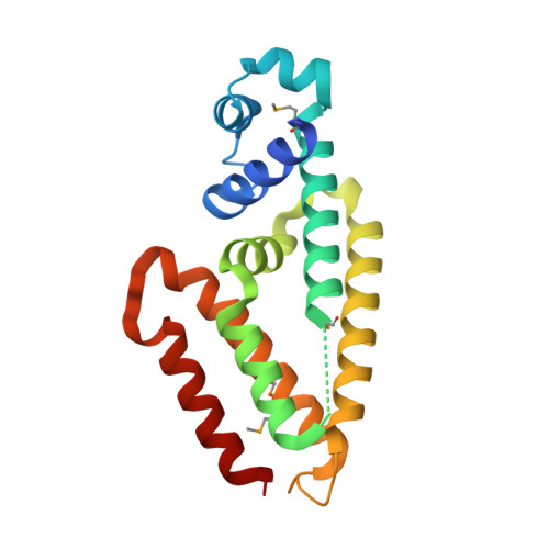
    Expression of the gene cluster icaADBC is necessary for biofilm production in Staphylococcus epidermidis. The ica operon is negatively controlled by the repressor IcaR. Here, the crystal structure of IcaR was determined and the refined structure revealed a homodimer comprising entirely alpha-helices, typical of the tetracycline repressor protein family for gene regulations. The N-terminal domain contains a conserved helix-turn-helix DNA-binding motif with some conformational variations, indicating flexibility in this region. The C-terminal domain shows a complementary surface charge distribution about the dyad axis, ideal for efficient and specific dimer formation. The results of the electrophoretic mobility shift assay and isothermal titration calorimetry suggested that a 28 bp core segment of the ica operator is implicated in the cooperative binding of two IcaR dimers on opposite sides of the duplex DNA. Computer modeling based on the known DNA-complex structure of QacR and site-specific mutagenesis experiments showed that direct protein-DNA interactions are mostly conserved, but with slight variations for recognizing the different sequences. By interfering with the binding of IcaR to DNA, aminoglycoside gentamicin and other antibiotics may activate the icaADBC genes and elicit biofilm production in S. epidermidis, and likely S. aureus, as a defense mechanism.


  • Organizational Affiliation
    • Institute of Biological Chemistry, Taipei, Taiwan.

Macromolecule Content 

  • Total Structure Weight: 47.02 kDa 
  • Atom Count: 3,888 
  • Modeled Residue Count: 357 
  • Deposited Residue Count: 384 
  • Unique protein chains: 1

Macromolecules

Find similar proteins by:|  3D Structure
Entity ID: 1
MoleculeChains  Sequence LengthOrganismDetailsImage
Biofilm operon icaABCD HTH-type negative transcriptional regulator icaR
A, B
192Staphylococcus epidermidis RP62AMutation(s): 4 
Gene Names: IcaR
UniProt
Find proteins for Q5HKQ1 (Staphylococcus epidermidis (strain ATCC 35984 / DSM 28319 / BCRC 17069 / CCUG 31568 / BM 3577 / RP62A))
Explore Q5HKQ1 
Go to UniProtKB:  Q5HKQ1
Entity Groups
Sequence Clusters30% Identity50% Identity70% Identity90% Identity95% Identity100% Identity
UniProt GroupQ5HKQ1
Sequence Annotations
Expand
Reference Sequence

Small Molecules

Modified Residues  1 Unique
IDChains TypeFormula2D DiagramParent
MSE
Query on MSE
A, B
L-PEPTIDE LINKINGC5 H11 N O2 SeMET

Experimental Data & Validation

Experimental Data

  • Method: X-RAY DIFFRACTION
  • Resolution: 1.33 Å
  • R-Value Free:  0.223 (Depositor), 0.218 (DCC) 
  • R-Value Work:  0.190 (Depositor), 0.190 (DCC) 
Space Group: P 1 21 1
Unit Cell:
Length ( Å )Angle ( ˚ )
a = 61.78α = 90
b = 50.7β = 97.09
c = 63.34γ = 90
Software Package:
Software NamePurpose
HKL-2000data collection
SOLVEphasing
CNSrefinement
HKL-2000data reduction
HKL-2000data scaling

Structure Validation

View Full Validation Report


Entry History 

Deposition Data

Revision History  (Full details and data files)

  • Version 1.0: 2008-02-05
    Type: Initial release
  • Version 1.1: 2011-07-13
    Changes: Source and taxonomy, Version format compliance
  • Version 1.2: 2021-11-10
    Changes: Database references, Derived calculations
  • Version 1.3: 2024-10-23
    Changes: Data collection, Structure summary